. 2006 Jun;26(11):4017-27.
doi: 10.1128/MCB.01904-05.

Protein phosphatase 2A stabilizes human securin, whose phosphorylated forms are degraded via the SCF ubiquitin ligase

Affiliations

Protein phosphatase 2A stabilizes human securin, whose phosphorylated forms are degraded via the SCF ubiquitin ligase

Ana M Gil-Bernabé et al. Mol Cell Biol. 2006 Jun.

Abstract

Sister chromatid segregation is triggered at the metaphase-to-anaphase transition by the activation of the protease separase. For most of the cell cycle, separase activity is kept in check by its association with the inhibitory chaperone securin. Activation of separase occurs at anaphase onset, when securin is targeted for destruction by the anaphase-promoting complex or cyclosome E3 ubiquitin protein ligase. This results in the release of the cohesins from chromosomes, which in turn allows the segregation of sister chromatids to opposite spindle poles. Here we show that human securin (hSecurin) forms a complex with enzymatically active protein phosphatase 2A (PP2A) and that it is a substrate of the phosphatase, both in vitro and in vivo. Treatment of cells with okadaic acid, a potent inhibitor of PP2A, results in various hyperphosphorylated forms of hSecurin which are extremely unstable, due to the action of the Skp1/Cul1/F-box protein complex ubiquitin ligase. We propose that PP2A regulates hSecurin levels by counteracting its phosphorylation, which promotes its degradation. Misregulation of this process may lead to the formation of tumors, in which overproduction of hSecurin is often observed.


Figures
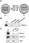
FIG. 1.
FIG. 1.
hSecurin interacts with the serine/threonine PP2A phosphatase. (A) hSec-E/A (residues 1 to 163 of hSecurin) interacts with p27 (residues 233 to 453 of PP2A-B55δ) in a yeast two-hybrid assay. Hf7c reporter strain was cotransformed with the indicated constructs. The interaction between the two-hybrid proteins is indicated by growth in the absence of histidine (dark gray patch). DBD, fusion with the DNA-binding domain of Gal4; AD, fusion with the activation domain of Gal4; none, empty vector. Snf1-Snf4 interaction was used as a positive control. (B) hSecurin interacts with PP2A-cs in vitro. Six-His fusion proteins were incubated with NP40 extracts from HeLa cells, purified on an Ni-NTA agarose column, and their association with PP2A-cs was determined by immunoblotting. RIIα is an unrelated protein. Lys, lysate from HeLa cells. (C) hSecurin binds to the PP2A complex in vivo. Anti-hSecurin and preimmune (PI) sera were used in immunoprecipitations from NP40 extracts of HeLa cells. The immunoprecipitates (IP) were probed for the presence of PP2A-B55α/δ, PP2A-cs, and hSecurin. Lys, lysate from HeLa cells.
FIG. 2.
FIG. 2.
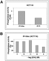
hSecurin immunoprecipitates contain phosphatase activity sensitive to okadaic acid. (A) hSecurin was immunoprecipitated from NP40 extracts of HCT116 cells. Preimmune serum (PI) was used as a control. Phosphatase activity level was standardized to that obtained from immunoprecipitates (IP) using the preimmune serum. (B) Immunoprecipitates of hSecurin from NP40 extracts of HCT116 cells were incubated with increasing concentrations of okadaic acid (OA). Activities are represented as a percentage of that observed in the control containing no OA (C). Similar findings were obtained in five separate experiments.
FIG. 3.
FIG. 3.
The phosphatase activity associated with hSecurin immunoprecipitates is dependent on the stage of the cell cycle and inhibited by okadaic acid. (A) hSecurin was immunoprecipitated from extracts of synchronized HeLa cells harvested at various cell cycle phases. Immunoprecipitates (IP) were probed for the presence of hSecurin and PP2A-cs. Lys, lysate from asynchronous HeLa cells. (B) Specific phosphatase activity of hSecurin immunoprecipitates, obtained as described for panel A, was measured in presence or absence of 1 nM okadaic acid (OA). Phosphatase activity was calculated as the amount of phosphate released from a synthetic phosphothreonine peptide. When indicated, OA was added to assay mixtures. Similar findings were obtained in five separate experiments.
FIG. 4.
FIG. 4.
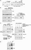
Specific phosphorylated forms of hSecurin, which are degraded by the proteasome in the presence of okadaic acid, are substrates of PP2A phosphatase. (A) Phosphorylated forms of hSecurin from nocodazole-treated cells are not substrates of PP2A phosphatase. hSecurin immunoprecipitates (IP) from nocodazole-treated HeLa cells were incubated with or without commercially obtained PP2A or lambda protein phosphatase (λ-PP) prior to Western blotting for detection of hSecurin. Asyn, lysate from asynchronous HeLa cells; NZ, lysate from nocodazole-treated HeLa cells. (B) In the upper panel, similarly to panel A, hSecurin immunoprecipitates from nocodazole-treated HeLa cells were incubated with commercial PP2A (+PP2A), B55δ immunocomplexes (+ IP B55δ), or both commercial PP2A and B55δ immunocomplexes, prior to Western blotting for detection of hSecurin. −, hSecurin immunoprecipitates from nocodazole-treated HeLa cells; Asyn, lysate from asynchronous HeLa cells; NZ, lysate from nocodazole-treated HeLa cells. (Lower panel) Cos-7 cells were transiently transfected with epitope-tagged hSecurin VSV (hSec VSV) prior to treatment with both okadaic acid (OA), and the proteasome and calpain inhibitor Ac-LLnL-CHO (LLnL). hSecurin immunoprecipitates from these cells were incubated either with commercial PP2A or B55δ immunocomplexes. Lysates and immunoprecipitates were Western blotted for hSecurin detection. Lane C, lysates from nontreated transfected Cos-7 cells; OA+LLnL, lysates from okadaic acid- and LLnL-treated transfected Cos-7 cells. (C) HeLa cells were treated with okadaic acid, LLnL, or both for 1 h 30 min prior to treatment with or without λ-PP for 30 min. Lysates were subsequently Western blotted for the detection of hSecurin. Lanes C, lysate from control untreated HeLa cells. (D) HeLa cells were transiently transfected with pCDNA3-hSec VSV along siRNA oligonucleotides against PP2A-cs α or nonspecific control oligonucleotides (mock) and collected after 48 h. LLnL was added 24 h after transfections. Extracts were Western blotted for PP2A-cs and hSecurin detection. −, lysate from nontransfected and nontreated HeLa cells. (E) Cos-7 cells were transiently transfected with hSec VSV prior to treatment with both okadaic acid and LLnL. hSecurin immunoprecipitates from these cells were incubated either with (+PP2A) or without (+Incub.) commercial PP2A. Lysates and immunoprecipitates were Western blotted for hSecurin detection. −, nonincubated hSecurin immunoprecipitates. Lane C, lysates from nontreated transfected Cos-7 cells; OA+LLnL, lysates from okadaic acid-and LLnL-treated transfected Cos-7 cells.
FIG. 5.
FIG. 5.
Phosphorylated forms of hSecurin induced by okadaic acid are not APC/C substrates but are degraded via the SCF E3 ubiquitin ligase. (A) Cos-7 cells transiently transfected with either hSecurin (hSec) or hSecurin with KEN-box and D-box mutations (hSec KAA-DM) were treated with okadaic acid (OA), LLnL, or both. Extracts, where indicated, were treated with λ-PP for 30 min. Western-blotted extracts were detected for hSecurin. Lanes C, lysates from nontreated transfected Cos-7 cells. (B) HeLa cells were transiently cotransfected with pSUPER-Cdh1 and pSUPER-Cdc20 or with pSUPER alone and harvested 48 h after transfection. When indicated, okadaic acid was added 1 h 30 min before cells were harvested. Extracts were Western blotted for the detection of Cdh1, Cdc20, hSecurin, and cyclin B. Note that the Western blot for Cdc20 shows three bands, the upper two (and most prominent) of which are cross-reactive and not specific to Cdc20. The right panel shows a Western blot of extracts which were treated with λ-PP. −, lysates from nontransfected HeLa cells. (C) In the left panels, Cos-7 cells transiently transfected with pCDNA3 or pCDNA3 Flag-Cul1(1-452) were treated either with (lanes OA) or without (lanes C) okadaic acid before harvesting. NP40 extracts were prepared and were Western blotted for Cul1, Flag-Cul1(1-452), and securin detection. Where indicated, extracts were treated with λ-PP. Expression of Cdc25A was also tested in Cos-7-transfected cells. (Right panels) Cos-7 cells transiently transfected with the indicated plasmids were treated either with (lanes OA) or without (lanes C) okadaic acid before harvesting. NP40 extracts were prepared and were Western blotted for Cul1, Flag-Cul1(1-452), hSec VSV, and EGFP detection. Where indicated, extracts were treated with λ-PP. (D) Anti-hSecurin and preimmune (PI) sera were used in immunoprecipitations from NP40 extracts of LLnL-treated Cos-7 cells. The immunoprecipitates (IP) were probed for the presence of Cul1 and hSecurin. Lys, lysate from LLnL-treated Cos-7 cells; IgG, the IgG heavy chains.
FIG. 6.
FIG. 6.
The PP2A phosphatase complex stabilizes hSecurin. (A) Cos-7 cells were transiently transfected with either empty vector (pECE) or pECE-B55δ(233-453). Endogenous securin levels (upper left panel) or cotransfected hSecurin VSV (pCDNA3-hSec VSV; upper right panel) was detected in immunoblotted lysates. EGFP (pEGFP-N1; right panel) was used as an internal control for transfection efficiency. (B) Cos-7 cells were transiently cotransfected with hSecurin VSV (hSec VSV) and either HA B55δ [pECE-B55δ(233-453)] or empty vector (pECE), and their expression was induced for 18 h. Cycloheximide (CHX; 50 μg/ml) was added to the media to inhibit protein synthesis, and cells were collected at the indicated times after addition of CHX. hSecurin VSV levels in cell lysates were detected by immunoblotting with anti-VSV monoclonal antibody. (C) HeLa cells were transiently transfected with siRNA oligonucleotides against PP2A-cs α or nonspecific control oligonucleotides (mock) and collected after 72 h. Western blots of extracts were analyzed for PP2A-cs, hSecurin, and cyclin B levels. −, extract from noninterfered HeLa cells. (D) HeLa cells were transiently transfected with the indicated plasmids along siRNA oligonucleotides against PP2A-cs α or nonspecific control oligonucleotides (mock) and collected after 48 h. Extracts were Western blotted for PP2A-cs and hSecurin detection.

Similar articles

Cited by

References

    1. Agarwal, R., and O. Cohen-Fix. 2002. Phosphorylation of the mitotic regulator Pds1/securin by Cdc28 is required for efficient nuclear localization of Esp1/separase. Genes Dev. 16:1371-1382. - PMC - PubMed
    1. Bernal, J. A., R. Luna, A. Espina, I. Lazaro, F. Ramos-Morales, F. Romero, C. Arias, A. Silva, M. Tortolero, and J. A. Pintor-Toro. 2002. Human securin interacts with p53 and modulates p53-mediated transcriptional activity and apoptosis. Nat. Genet. 32:306-311. - PubMed
    1. Bhatia, K., K. Huppi, G. Spangler, D. Siwarski, R. Iyer, and I. Magrath. 1993. Point mutations in the c-Myc transactivation domain are common in Burkitt's lymphoma and mouse plasmacytomas. Nat. Genet. 5:56-61. - PubMed
    1. Breeden, L., and K. Nasmyth. 1985. Regulation of the yeast HO gene. Quant. Biol. 50:643-650. - PubMed
    1. Brummelkamp, T. R., R. Bernards, and R. Agami. 2002. A system for stable expression of short interfering RNAs in mammalian cells. Science 296:550-553. - PubMed

Publication types

Substances

LinkOut - more resources