. 2006 Jun;26(11):4041-51.
doi: 10.1128/MCB.01868-05.

Insulin-like growth factor I controls a mutually exclusive association of RACK1 with protein phosphatase 2A and beta1 integrin to promote cell migration

Affiliations

Insulin-like growth factor I controls a mutually exclusive association of RACK1 with protein phosphatase 2A and beta1 integrin to promote cell migration

Patrick A Kiely et al. Mol Cell Biol. 2006 Jun.

Abstract

The WD repeat scaffolding protein RACK1 can mediate integration of the insulin-like growth factor I receptor (IGF-IR) and integrin signaling in transformed cells. To address the mechanism of RACK1 function, we searched for regulatory proteins that associate with RACK1 in an IGF-I-dependent manner. The serine threonine phosphatase protein phosphatase 2A (PP2A) was found associated with RACK1 in serum-starved cells, and it dissociated immediately upon stimulation with IGF-I. This dissociation of PP2A from RACK1 and an IGF-I-mediated decrease in cellular PP2A activity did not occur in cells expressing either the serine 1248 or tyrosine 1250/1251 mutants of the IGF-IR that do not interact with RACK1. Recombinant RACK1 could bind to PP2A in vitro and restore phosphatase activity to PP2A from IGF-I-stimulated cells. Ligation of integrins with fibronectin or Matrigel was sufficient to facilitate IGF-I-mediated dissociation of PP2A from RACK1 and also to recruit beta1 integrin as PP2A dissociated. By using TAT-fused N-terminal and C-terminal deletion mutants of RACK1, we determined that both PP2A and beta1 integrin interact in the C terminus of RACK1 within WD repeats 4 to 7. This suggests that integrin ligation displaces PP2A from RACK1. MCF-7 cells overexpressing RACK1 exhibited enhanced motility, which could be reversed by the PP2A inhibitor okadaic acid. Small interfering RNA-mediated suppression of RACK1 also decreased the migratory capacity of DU145 cells. Taken together, our findings indicate that RACK1 enhances IGF-I-mediated cell migration through its ability to exclusively associate with either beta1 integrin or PP2A in a complex at the IGF-IR.


Figures

FIG. 1.
FIG. 1.
RACK1 interacts with PP2A and dissociates upon IGF stimulation. MCF-7, PC-3, or DU145 cells were starved and stimulated with IGF-I for the indicated times. Cell lysates were immunoprecipitated with anti-RACK1 antibody, followed by Western blotting with anti-PP2A C antibody. The presence of RACK1 in the immunoprecipitate was confirmed using anti-RACK1 antibody on the same blot. (A) MCF-7 cells. (B) PC-3 and DU145 cells. (C) Purified PP2A (0.5 μg) was incubated with column-bound MBP or MBP-RACK1. After the proteins were washed extensively, they were eluted with maltose and assessed by Western blotting for associated PP2A. MCF-7 cell lysate was a control for Western blotting. The results from two separate preparations of MBP-RACK1 are shown along with a Coomassie-stained gel of the bacterial crude lysate from which MBP and MBP-RACK1 were affinity purified.
FIG. 2.
FIG. 2.
The association of RACK1 with IGF-IR regulates the association of RACK1 with PP2A. (A) R cells were transiently transfected with plasmids encoding full-length WT IGF-IR or the Y1250/1251F and S1248A mutants. IGF-IR was immunoprecipitated, followed by Western blotting with anti-RACK1 antibody to detect associated endogenous RACK1. The blot was reprobed with anti-IGF-IR antibody to demonstrate expression of IGF-IR. (B) WT, Y1250/1251F, or S1248A IGF-IR was transiently expressed into R cells. Endogenous RACK1 was immunoprecipitated with anti-RACK1 antibody, followed by Western blotting with anti-PP2A C antibody. The membrane was reprobed with anti-RACK1 antibody to confirm that comparable levels of RACK1 were immunoprecipitated in each sample. A lysate control from the experiment was included, showing expression of each transfected IGF-IR. (C, left panel) MCF-7 cells were starved for 4 h and stimulated with IGF-I. Cell lysates were prepared and PP2A immunoprecipitated prior to measurement of PP2A activity, using p-NPP as a substrate. Okadaic acid (OA) (1 nM) was added where indicated, 1 h prior to cell lysis. Data are presented from triplicate samples, where PP2A activity in cells not stimulated with IGF-I is represented as 100%. (C, right panel) R cells transiently transfected with plasmids encoding full-length IGF-IR or the Y1250/1251F mutant IGF-IR were serum starved for 4 h and stimulated with IGF-I for the indicated times. Cell lysates were prepared and PP2A immunoprecipitated prior to assay of PP2A activity. Data from triplicate samples are presented, with 100% activity representing that measured in cells expressing WT IGF-IR not stimulated with IGF-I.
FIG. 3.
FIG. 3.
Adhesion signals are required for IGF-I-mediated dissociation of PP2A from RACK1. (A) Cell lysates were prepared from MCF-7 cells that were serum starved under adherent conditions or while maintained in nonadherent cultures for 4 h. The cells were then stimulated with IGF-I, and RACK1 was immunoprecipitated, followed by Western blotting with anti-PP2A C antibody. The membrane was reprobed with anti-RACK1 antibody. (B) Cell lysates were prepared from adherent MCF-7 cells that had been serum starved for 4 h and from nonadherent MCF-7 cells that had been starved and stimulated with IGF-I. PP2A was immunoprecipitated and assayed for activity, using p-NPP as a substrate. Data are presented for triplicate samples, where 100% activity represents that measured in adherent cells not stimulated with IGF-I. (C and D) Cell lysates were prepared from MCF-7 cells that were serum starved for 4 h or that were serum starved in nonadherent cultures for 4 h in the presence of (C) fibronectin (5 μg/ml) or (D) Matrigel (5%) prior to stimulation with IGF-I. RACK1 was immunoprecipitated, followed by Western blot analysis to detect associated β1 integrin or PP2A. The blot was also probed with anti-RACK1 antibody.
FIG. 4.
FIG. 4.
Both PP2A and β1 integrin interact with WD repeats 4 to 7 of RACK1. (A) Model illustrating the N-terminal fragment of RACK1, amino acids 1 to 180 (RACK1ΔC), or the C-terminal fragment of RACK1, amino acids 138 to 317 (RACK1ΔN), both of which have WD4 and can bind endogenous RACK1. (B) MCF-7 cells were transduced with the TAT fusion proteins TAT-RACK1, RACK1ΔC, and RACK1ΔN for 2 h at 37°C. TAT-RACK1 proteins were immunoprecipitated from cells by using anti-HA antibodies and probed by Western blotting for associated endogenous RACK1. (C) Immunoprecipitated TAT-RACK1, RACK1ΔC, and RACK1ΔN were investigated for association with PP2A and β1 integrin by Western blotting. (D) MCF-7 cells were transduced with the TAT fusion proteins, serum starved for 3 h, and stimulated with IGF-I for the indicated times. TAT-RACK1, RACK1ΔC, and RACK1ΔN were immunoprecipitated from cell lysates by using anti-HA antibodies and then investigated for associated PP2A and β1 integrin by Western blotting. FL, full length.
FIG. 5.
FIG. 5.
RACK1 restores PP2A activity from IGF-I-stimulated cells in vitro. RACK1 was cleaved from MBP, using factor Xa. PP2A was immunoprecipitated from serum-starved or IGF-I-stimulated MCF-7 cells and either cleaved RACK1 or MBP was incubated with bead-bound immune complexes or with purified PP2A for 45 min prior to assay of PP2A activity. Data are presented from triplicate samples, where PP2A activity in MCF-7 cells not stimulated with IGF-I is represented as 100%. A sample from MCF-7 cells where okadaic acid (OA) was added prior to cell lysis was also included as a control.
FIG. 6.
FIG. 6.
RACK1 enhances MCF-7 cell motility and migration toward IGF-I. (A) MCF-7/Neo and MCF-7/HARACK1 cells were grown to 60% confluence in medium supplemented with 10% fetal calf serum. A wound was scored in each culture and cells allowed to migrate for 24 h. Cells were then stained with Giemsa and photographed at a magnification of ×10. A representative of multiple similar fields is presented for each condition. (B) The migratory potentials of MCF-7/ Neo and MCF-7/HARACK1 cells were compared in Transwell assays. Cells were plated at 50,000 cells/chamber and allowed to migrate toward IGF-I for 24 h. Cells were fixed by using methanol before they were stained with crystal violet and examined under a magnification of ×10. Expression of RACK1 in each cell line tested compared with that in MCF-7 cells is shown in Western blots in right-hand panels. (C) MCF-7/Neo and MCF-7/HARACK1 cells were starved and stimulated with IGF-I. Cell lysates were immunoprecipitated with anti-β1 integrin antibody, followed by Western blotting with anti-RACK1 antibody. The presence of IGF-IR in the immunoprecipitate was confirmed by using anti-IGF-IR antibody on the same blot. A lysate control is included to compare levels of RACK1 in the cells. (D) IGF-IR was immunoprecipitated from MCF-7, MCF-7/Neo, and MCF-7/HARACK1 cells, and RACK1 was detected in the immunoprecipitate by probing the blot with anti-RACK1 antibody. (E) Lysates from MCF-7/Neo or MCF-7/RACK1 cells were assessed by Western blotting for FAK phosphorylation by using anti-phospho-FAK (Y397) antibody. The blot was stripped and reprobed for FAK and actin as loading controls.
FIG. 6.
FIG. 6.
RACK1 enhances MCF-7 cell motility and migration toward IGF-I. (A) MCF-7/Neo and MCF-7/HARACK1 cells were grown to 60% confluence in medium supplemented with 10% fetal calf serum. A wound was scored in each culture and cells allowed to migrate for 24 h. Cells were then stained with Giemsa and photographed at a magnification of ×10. A representative of multiple similar fields is presented for each condition. (B) The migratory potentials of MCF-7/ Neo and MCF-7/HARACK1 cells were compared in Transwell assays. Cells were plated at 50,000 cells/chamber and allowed to migrate toward IGF-I for 24 h. Cells were fixed by using methanol before they were stained with crystal violet and examined under a magnification of ×10. Expression of RACK1 in each cell line tested compared with that in MCF-7 cells is shown in Western blots in right-hand panels. (C) MCF-7/Neo and MCF-7/HARACK1 cells were starved and stimulated with IGF-I. Cell lysates were immunoprecipitated with anti-β1 integrin antibody, followed by Western blotting with anti-RACK1 antibody. The presence of IGF-IR in the immunoprecipitate was confirmed by using anti-IGF-IR antibody on the same blot. A lysate control is included to compare levels of RACK1 in the cells. (D) IGF-IR was immunoprecipitated from MCF-7, MCF-7/Neo, and MCF-7/HARACK1 cells, and RACK1 was detected in the immunoprecipitate by probing the blot with anti-RACK1 antibody. (E) Lysates from MCF-7/Neo or MCF-7/RACK1 cells were assessed by Western blotting for FAK phosphorylation by using anti-phospho-FAK (Y397) antibody. The blot was stripped and reprobed for FAK and actin as loading controls.
FIG. 7.
FIG. 7.
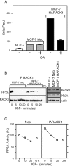
Okadaic acid reverses the enhanced motility of MCF-7/RACK1 cells. (A) MCF-7/Neo and MCF-7/HARACK1 cells to IGF-I were compared for migratory potential in Transwell assays. Cells were starved and incubated with or without okadaic acid (OA) at 1 nM, seeded at 50,000 cells/chamber, and allowed to migrate toward IGF-I for 16 h. Cells were fixed by using methanol before they were stained with crystal violet and examined under a magnification of ×10. The data are presented as the number of cells/well for triplicate wells of each culture condition. MCF-7 cells are included as a control. (B) MCF-7/Neo and MCF-7/HARACK1 cells were starved and stimulated with IGF-I for the indicated times. Cell lysates were immunoprecipitated (IP) with anti-RACK1 antibody, followed by Western blotting with anti-PP2A C antibody. The presence of RACK1 in the immunoprecipitates was confirmed by using anti-RACK1 antibody on the same blot. Levels of each protein in cell lysates are shown in the right-hand panel. (C) PP2A activities in MCF-7/Neo (Neo) and MCF-7/HARACK1 (HARACK1) cells were assayed, using p-NPP as a substrate. The data are presented from triplicate samples, where 100% activity is represented by Neo cells not stimulated with IGF-I.
FIG. 8.
FIG. 8.
RACK1 siRNA ablates IGF-I-mediated regulation of PP2A activity and cell migration. (A) MCF-7 cells transfected with two different siRNA oligonucleotides directed to RACK1 (siRNA1 and siRNA2) or a scrambled oligonucleotide (control) were assessed for RACK1 expression by Western blotting (upper panels). PP2A was immunoprecipitated from cells stimulated or not stimulated with IGF-I and was assayed for phosphatase activity. Data from triplicate samples are presented, with 100% activity representing that measured in MCF-7 cells transfected with a negative control. (B) To investigate the effects of siRNA RACK1 on cell migration, DU145 cells were transfected with siRNA1 or a control oligonucleotide (10 μM). Expression of RACK1 was assessed by Western blotting (upper panel). Cells were assessed for migration toward serum in Transwell assays at 24 h posttransfection. Cells were fixed by using methanol before they were stained with crystal violet and counted under a magnification of ×10.
FIG. 9.
FIG. 9.
Model to illustrate actions of RACK1 in regulating recruitment of β1 integrin and PP2A to control cell migration. The left panel represents serum-starved cells in which RACK1 is associated with the IGF-IR and PP2A. FAK phosphorylation and PP2A activity are high, and there is no cell migration. The right panel represents cells stimulated with IGF-I. PP2A is rapidly released from RACK1, and β1 integrin is recruited to RACK1, resulting in a complex that includes the IGF-IR and β1 integrin. PP2A activity is transiently decreased, FAK phosphorylation is decreased, and cell migration is stimulated.

Similar articles

Cited by

References

    1. Allan, G. J., D. J. Flint, and K. Patel. 2001. Insulin-like growth factor axis during embryonic development. Reproduction 122:31-39. - PubMed
    1. Beaulieu, J. M., T. D. Sotnikova, S. Marion, R. J. Lefkowitz, R. R. Gainetdinov, and M. G. Caron. 2005. An Akt/beta-arrestin 2/PP2A signaling complex mediates dopaminergic neurotransmission and behavior. Cell 122:261-273. - PubMed
    1. Berns, H., R. Humar, B. Hengerer, F. N. Kiefer, and E. J. Battegay. 2000. RACK1 is up-regulated in angiogenesis and human carcinomas. FASEB J. 14:2549-2558. - PubMed
    1. Boudreau, R. T., R. Garduno, and T. J. Lin. 2002. Protein phosphatase 2A and protein kinase Calpha are physically associated and are involved in Pseudomonas aeruginosa-induced interleukin 6 production by mast cells. J. Biol. Chem. 277:5322-5329. - PubMed
    1. Buensuceso, C. S., D. Woodside, J. L. Huff, G. E. Plopper, and T. E. O'Toole. 2001. The WD protein Rack1 mediates protein kinase C and integrin-dependent cell migration. J. Cell Sci. 114:1691-1698. - PubMed

Publication types

MeSH terms

LinkOut - more resources