. 2006 Jun;26(11):4149-60.
doi: 10.1128/MCB.01932-05.

T-cell protein tyrosine phosphatase (Tcptp) is a negative regulator of colony-stimulating factor 1 signaling and macrophage differentiation

Affiliations

T-cell protein tyrosine phosphatase (Tcptp) is a negative regulator of colony-stimulating factor 1 signaling and macrophage differentiation

Paul D Simoncic et al. Mol Cell Biol. 2006 Jun.

Abstract

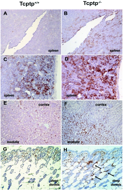
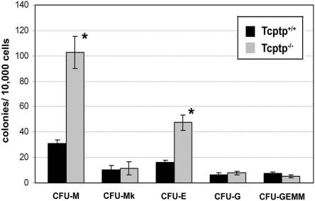
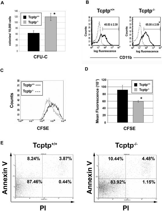
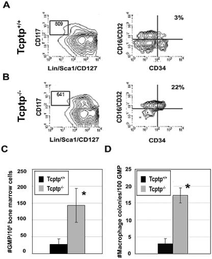
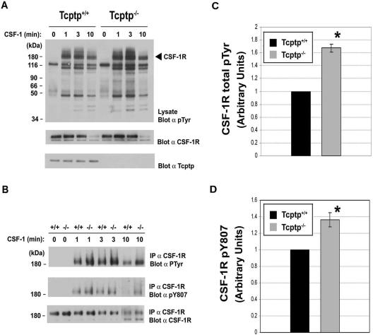
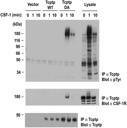
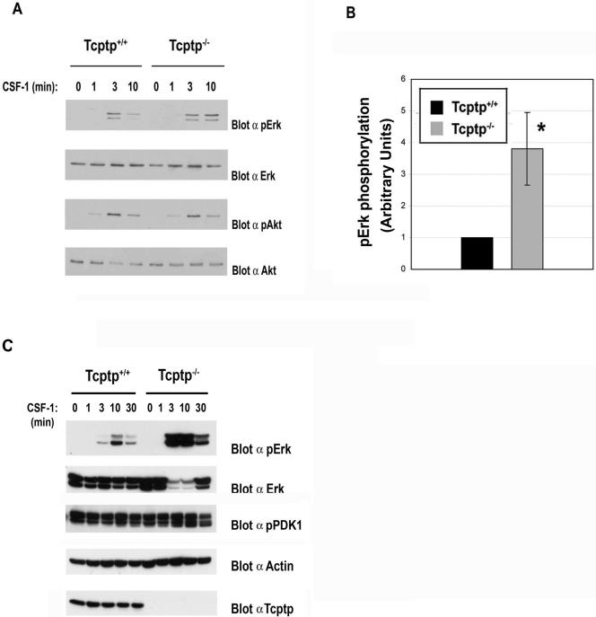
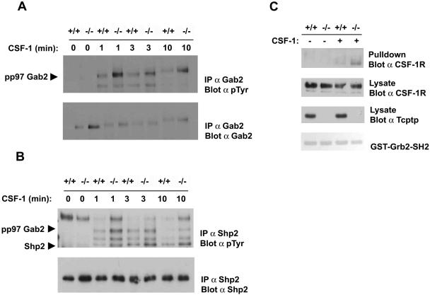
Mice null for the T-cell protein tyrosine phosphatase (Tcptp-/-) die shortly after birth due to complications arising from the development of a systemic inflammatory disease. It was originally reported that Tcptp-/- mice have increased numbers of macrophages in the spleen; however, the mechanism underlying the aberrant growth and differentiation of macrophages in Tcptp-/- mice is not known. We have identified Tcptp as an important regulator of colony-stimulating factor 1 (CSF-1) signaling and mononuclear phagocyte development. The number of CSF-1-dependent CFU is increased in Tcptp-/- bone marrow. Tcptp-/- mice also have increased numbers of granulocyte-macrophage precursors (GMP), and these Tcptp-/- GMP yield more macrophage colonies in response to CSF-1 relative to wild-type cells. Furthermore, we have identified the CSF-1 receptor (CSF-1R) as a physiological target of Tcptp through substrate-trapping experiments and its hyperphosphorylation in Tcptp-/- macrophages. Tcptp-/- macrophages also have increased tyrosine phosphorylation and recruitment of a Grb2/Gab2/Shp2 complex to the CSF-1R and enhanced activation of Erk after CSF-1 stimulation, which are important molecular events in CSF-1-induced differentiation. These data implicate Tcptp as a critical regulator of CSF-1 signaling and mononuclear phagocyte development in hematopoiesis.


Figures

FIG. 1.
FIG. 1.
Tissues from Tcptp−/− mice are infiltrated with F4/80+ macrophages. Tissue sections from Tcptp+/+ (A, C, E, and G) or Tcptp−/− (B, D, F, and H) mice were stained with an anti-F4/80 antibody to detect tissue macrophages. Sections from spleen (A, B, C, and D), kidney (E and F), and skin (G and H) are shown. Panels A, B, E, and F were photographed at an original magnification of ×20, while panels C, D, G, and H were photographed at an original magnification of ×40. For each panel, a minimum of three mice of each genotype were analyzed. Arrows indicate staining of deep dermal macrophages.
FIG. 2.
FIG. 2.
Increased numbers of macrophage precursors in Tcptp−/− bone marrow. Tcptp+/+ or Tcptp−/− bone marrow cells (1 × 10⁴) were plated in methylcellulose containing IL-3, SCF, IL-6, and Epo. At day 12, colonies were scored under high magnification to determine colony constitution and size (means ± SD [error bars]; n = 7; *, P < 0.01).
FIG. 3.
FIG. 3.
Increased mononuclear phagocytic precursors in Tcptp−/− bone marrow. (A) Macrophage colonies derived from CFU-C in Tcptp+/+ or Tcptp−/− bone marrow cells (1 × 10⁴) cultured in the presence of 100 ng/ml CSF-1. Colonies were scored at day 7 (n = 6; means ± SD [error bars]; *, P < 0.01). (B) Bone marrow cells from Tcptp+/+ or Tcptp−/− mice were cultured for 3 days in the presence of IL-3 and CSF-1 to allow proliferation and differentiation of multipotent progenitors to nonadherent mononuclear phagocytic precursors. Cells were stained with anti-CD11b-FITC antibodies (solid line) to determine the percentage of mononuclear phagocytic precursors in both cell populations. Staining with an isotype-matched control antibody is shown as a dashed line (means ± SD [error bars]; n = 4, *, P < 0.01). (C) Increased proliferation of Tcptp−/− committed mononuclear phagocyte precursor cells. Bone marrow cells were cultured as described for panel A to generate committed mononuclear phagocytes, and cells were labeled with CFSE and allowed to proliferate for 3 days. CFSE fluorescence was measured by flow cytometry, and cell populations were plotted as histograms. (D) Geometric means from histograms in panel C were plotted to quantitate CFSE dilution. Data were analyzed by an unpaired, two-tailed Student t test (means ± SD [error bars]; n = 3; *, P < 0.05). (E) Annexin V-propidium iodide staining of apoptotic cells in the mononuclear phagocyte cultures used in CFSE proliferation assay.
FIG. 4.
FIG. 4.
Increased sensitivity of GMP from Tcptp−/− bone marrow to CSF-1. GMP from Tcptp+/+ (A) and Tcptp−/− (B) bone marrow were purified by FACS. These cells are defined as Lin/Sca1/CD127(IL-7Rα), CD117(c-KIT)+, FcR+, and CD34+. (C) Graphical representation of the number of GMP in Tcptp+/+ and Tcptp−/− per 106 bone marrow cells. Error bars indicate standard deviations. (n = 4; *, P < 0.01). (D) Colonies derived from 100 GMP in clonogenic assays with 100 ng/ml CSF-1. (means ± SD [error bars]; n = 4, *, P < 0.01).
FIG. 5.
FIG. 5.
The CSF-1R is hyperphosphorylated in Tcptp−/− macrophages. (A) BMDMs from Tcptp+/+ or Tcptp−/− mice were stimulated with 100 ng/ml CSF-1 for the indicated time points. Equal amounts of lysates were resolved by SDS-PAGE and analyzed by Western blotting with an antiphosphotyrosine antibody (top panel). The blot was reprobed with anti-CSF-1R antibodies (middle panel) and anti-Tcptp antibodies (bottom panel) to demonstrate the correct genotype of the cells. (B) The CSF-1R was immunoprecipitated from Tcptp+/+ or Tcptp−/− BMDMs stimulated with 100 ng/ml CSF-1 as indicated. The immunoprecipitates were resolved by SDS-PAGE and analyzed by Western blotting with antiphosphotyrosine (top panel), anti-CSF-1R phospho-Y807 (middle panel), and anti-CSF-1R (bottom panel) antibodies. (C and D) Graphical representation of total tyrosine phosphorylation of the CSF-1R (C) or phosphorylation of Y807 (D) in Tcptp+/+ or Tcptp−/− BMDMs at 10 min after CSF-1 stimulation. Western blots were quantitated by densitometry. Data were analyzed by an unpaired, two-tailed Student t test with the results of the wild-type set to 1 (means ± SD [error bars]; n = 3; *, P < 0.05).
FIG. 6.
FIG. 6.
The CSF-1R coimmunoprecipitates with Tcptp substrate-trapping mutants in vivo. FD-Fms cells were transfected with pEF-Tcptp-WT, pEF-Tcptp-DA, or empty vector, and then stimulated with 100 ng/ml CSF-1. Tcptp immunoprecipitates were analyzed by Western blotting with antiphosphotyrosine antibodies to reveal coimmunoprecipitating substrates (top panel). Immunoprecipitates were then blotted with anti-CSF-1R antibodies (middle panel) and anti-Tcptp antibodies (bottom panel) to ensure overexpression of the indicated constructs. The three right lanes contain whole-cell lysates of vector transfected cells to demonstrate the specificity of Tcptp for the tyrosine phosphorylated CSF-1R.
FIG. 7.
FIG. 7.
Tcptp−/− macrophages have increased Erk activation. (A) Lysates from Tcptp+/+ or Tcptp−/− BMDMs stimulated with 100 ng/ml CSF-1 were analyzed by Western blotting with anti-phospho-Erk and anti-phospho-Akt antibodies. (B) Graphical representation of total Erk1/2 TEY phosphorylation in Tcptp+/+ or Tcptp−/− BMDMs at 10 min CSF-1 stimulation. Western blots were quantitated by densitometry. Data were analyzed by an unpaired, two-tailed Student t test with the results of the wild-type set to 1 (means ± SD [error bars]; n = 3, *, P < 0.05). (C) Increased Erk phosphorylation after CSF-1 sitmulation in Tcptp−/− BMDMs is not prolonged. PDK1 activity in lysates from Tcptp+/+ or Tcptp−/− BMDMs stimulated with 100 ng/ml CSF-1 was also analyzed by Western blotting with anti-phospho-PDK1 antibodies.
FIG. 8.
FIG. 8.
Increased recruitment and tyrosine phosphorylation of a Grb2/Gab2/Shp2 complex after CSF-1 stimulation in Tcptp−/− macrophages. (A) Gab2 was immunoprecipitated from CSF-1-stimulated Tcptp+/+ or Tcptp−/− BMDMs. Protein complexes were resolved by SDS-PAGE and analyzed by antiphosphotyrosine blotting and then by blotting for total Gab2 protein. (B) Anti-Shp2 immunoprecipitates from CSF-1-stimulated Tcptp+/+ or Tcptp−/− BMDMs were analyzed by antiphosphotyrosine immunoblotting as described for panel A. (C) Lysates from unstimulated and CSF-1-stimulated Tcptp+/+ or Tcptp−/− BMDMs were incubated with a GST-Grb2-SH2 domain fusion protein. The complexes were resolved by SDS-PAGE and analyzed by Western blotting with an anti-CSF-1R antibody. Input lysates from the experiment were blotted with an anti-CSF-1R antibody to ensure equal amounts of the CSF-1R were used and with an anti-Tcptp antibody to ensure the genotype of the cells used in the experiment.

Similar articles

Cited by

References

    1. Alberola-Ila, J., and G. Hernandez-Hoyos. 2003. The Ras/MAPK cascade and the control of positive selection. Immunol. Rev. 191:79-96. - PubMed
    1. Alessi, D. R., S. R. James, C. P. Downes, A. B. Holmes, P. R. Gaffney, C. B. Reese, and P. Cohen. 1997. Characterization of a 3-phosphoinositide-dependent protein kinase which phosphorylates and activates protein kinase Bα. Curr. Biol. 7:261-269. - PubMed
    1. Austyn, J. M., and S. Gordon. 1981. F4/80, a monoclonal antibody directed specifically against the mouse macrophage. Eur. J. Immunol. 11:805-815. - PubMed
    1. Bartelmez, S. H., T. R. Bradley, I. Bertoncello, D. Y. Mochizuki, R. J. Tushinski, E. R. Stanley, A. J. Hapel, I. G. Young, A. B. Kriegler, and G. S. Hodgson. 1989. Interleukin 1 plus interleukin 3 plus colony-stimulating factor 1 are essential for clonal proliferation of primitive myeloid bone marrow cells. Exp. Hematol. 17:240-245. - PubMed
    1. Berg, K. L., K. A. Siminovitch, and E. R. Stanley. 1999. SHP-1 regulation of p62(DOK) tyrosine phosphorylation in macrophages. J. Biol. Chem. 274:35855-35865. - PubMed

Publication types

MeSH terms

Substances