. 2006 Jun;26(11):4201-13.
doi: 10.1128/MCB.01612-05.

Focal adhesion kinase controls cellular levels of p27/Kip1 and p21/Cip1 through Skp2-dependent and -independent mechanisms

Affiliations

Focal adhesion kinase controls cellular levels of p27/Kip1 and p21/Cip1 through Skp2-dependent and -independent mechanisms

Patrick Bryant et al. Mol Cell Biol. 2006 Jun.

Abstract

Endothelial cell proliferation is a critical step in angiogenesis and requires a coordinated response to soluble growth factors and the extracellular matrix. As focal adhesion kinase (FAK) integrates signals from both adhesion events and growth factor stimulation, we investigated its role in endothelial cell proliferation. Expression of a dominant-negative FAK protein, FAK-related nonkinase (FRNK), impaired phosphorylation of FAK and blocked DNA synthesis in response to multiple angiogenic stimuli. These results coincided with elevated cyclin-dependent kinase inhibitors (CDKIs) p21/Cip and p27/Kip, as a consequence of impaired degradation. FRNK inhibited the expression of Skp2, an F-box protein that targets CDKIs, by inhibiting mitogen-induced mRNA. The FAK-regulated degradation of p27/Kip was Skp2 dependent, while levels of p21/Cip were regulated independent of Skp2. Skp2 is required for endothelial cell proliferation as a consequence of degrading p27. Finally, knockdown of both p21 and p27 in FRNK-expressing cells completely restored mitogen-induced endothelial cell proliferation. These data demonstrate a critical role for FAK in the regulation of CDKIs through two independent mechanisms: Skp2 dependent and Skp2 independent. They also provide important insights into the requirement of focal adhesion kinase for normal vascular development and reveal novel regulatory control points for angiogenesis.


Figures
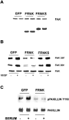
FIG. 1.
FIG. 1.
FRNK expression inhibits FAK and paxillin phosphorylation. HUVECs were transduced with Ad.GFP, Ad.FRNK, or Ad. FRNKS for 16 h in serum-free MCDB-131 and then treated with 50 ng/ml VEGF for 10 min, and whole cell lysates were collected and analyzed by sodium dodecyl sulfate-polyacrylamide gel electrophoresis and immunoblotting using antibodies to total FAK (A) and phosphorylation-specific antibodies to Y397 and Y861 (B). HUVECs were transduced with Ad.GFP or Ad.GFP-FRNK for 16 h in serum-free MCDB-131 and then treated with serum for 10 min and analyzed with total paxillin and Y118 paxillin antibodies (C). pPaxillin, phosphorylated paxillin.
FIG. 2.
FIG. 2.
FRNK expression inhibits mitogen-induced endothelial cell proliferation. HUVECs (A, B, and D) or HDMVECs were serum starved and transduced with Ad.GFP, Ad.FRNK, or Ad.FRNKS prior to stimulation. The cells were then incubated with 50 ng/ml VEGF (or complete growth medium; Fig. 3D) for 16 h. Cells were then pulsed with [3H]thymidine (H3 counts; A, C, and D) for an additional 3 h prior to scintillation counting. Cells pulsed with BrdU were visualized with an anti-BrdU antibody. BrdU-positive cells were quantified, and the data were graphed as the percentage of positive cells compared to total cell number (B).
FIG. 3.
FIG. 3.
Expression of FRNK does not induce apoptosis or disrupt cell spreading. (A) HUVECs were transduced with Ad.GFP or Ad.FRNK-GFP and analyzed at 48 h postinfection for the presence of caspase activity using a fluorescent substrate (Apo-ONE; Promega). Apoptosis was induced with a combination of TNF and alpha interferon (INF-α), as a positive control. RFLU, relative fluorescence units. (B) Adherent HUVECs were plated and transduced with Ad.GFP, Ad.FRNK-GFP, or Ad.FRNKS for 16 h under serum-free conditions. The total average cell surface area was calculated using Image-Pro Plus (Media Cybernetics) software. (C) Adherent HUVECs were placed in serum-free MCDB-131, infected with Ad.GFP or Ad.GFP-FRNK for 16 h, and then washed with 1× PBS and fixed with 3.7% formaldehyde. Cells were stained with Texas red-phalloidin and DAPI, and images of GFP, actin stress fibers, and nuclear staining were captured under ×100 magnification. White arrows indicate localization of GFP-FRNK at focal adhesion sites.
FIG. 4.
FIG. 4.
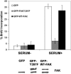
Expression of an Y397F mutant of FAK also inhibits endothelial cell proliferation. HUVECs were serum starved and transduced with Ad.GFP, Ad.GFP-Y397F-FAK, or Ad.GFP-WT-FAK prior to stimulation. The cells were then incubated with complete growth medium for 16 h. Cells were then pulsed with BrdU and visualized with an anti-BrdU antibody and recorded with a fluorescence digital camera. BrdU-positive cells were quantified, and the data were graphed as the percentage of positive cells compared to total cell number.
FIG. 5.
FIG. 5.
Expression of FRNK does not inhibit ERK signaling but does prevent serum-induced degradation of the CDKIs p27 and p21. (A) Serum-starved HUVECs were transduced with Ad.GFP, Ad.FRNK, or Ad.FRNKS and then stimulated with complete growth medium for 16 h and probed with antibodies specific for pJNK, pERK, pElk, and cyclin D1. (B) Serum-starved HUVECs were transduced with Ad.GFP or Ad.GFP-FRNK prior to stimulation with complete growth medium for 16 h. Cell lysates were made after 16 h and probed with antibodies specific for FAK Y397, p27, p21, pRb S795, and ERK2.
FIG. 6.
FIG. 6.
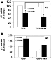
FRNK does not alter the mRNA expression of p27 or p21. Real-time PCR was performed on HUVECs expressing GFP (control) or GFP-FRNK for 16 h in serum-free media, at which time complete growth medium was added for an additional 16 h to the cells as indicated. The mRNA was then reversed transcribed and analyzed by quantitative real-time PCR using primers specific to p27 (A) and p21 (B) with a Bio-Rad real-time PCR machine (A and B). Data are normalized to the control value and are reported as percentage of increase ± standard error of three experiments. *, significantly different from serum-free paired controls (P < 0.05). NS, nonsignificant difference from serum-stimulated GFP controls.
FIG. 7.
FIG. 7.
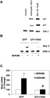
FRNK inhibits mitogen-induced expression of Skp2 protein and mRNA. Asynchronously growing HUVECs were treated with the proteasome inhibitors lactacystin and MG-132 as shown for 24 h, and then lysates were analyzed with antibodies specific to p27 and p21. Western blot analysis was then performed to analyze the levels of Skp2 protein expression after the addition of complete growth medium for 16 h (B). HUVECs were treated as in panel B, except mRNA was collected instead of protein using an RNeasy kit (QIAGEN). The mRNA was then reversed transcribed, and real-time PCR was performed using primers to Skp2 (C). Data are plotted as mean ± standard error. *, statistically significant difference from GFP; #, statistically significant difference from GFP treated with serum (P < 0.05) (n = 5).
FIG. 8.
FIG. 8.
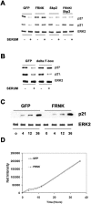
Skp2 regulates the FAK-dependent degradation of p27 but not p21. HUVECs were transduced with Ad.GFP, Ad.GFP-FRNK, Ad.WT-Skp2, or Ad.GFP-FRNK and Ad.WT-Skp2 combined in serum-free MCDB-131. The HUVECs were then treated with serum for 16 h and probed for p27, p21, and ERK (A). HUVECs were transduced with Ad.GFP or Ad.ΔF-box Skp2 treated the same as in panel A and then probed for p27, p21, and ERK2 (B). HUVECs in complete growth medium were transduced with Ad.GFP or Ad.GFP-FRNK for 6 h, treated with 10 μM lactacystin for the indicated times, and then lysed and probed for p21 protein levels (C). The net intensity of each band was quantified using a Kodak digital camera along with densitometry software (D).
FIG. 9.
FIG. 9.
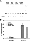
Skp2 regulates endothelial cell proliferation through modulation of p27. HUVECs were electroporated with 150 to 200 ng of siRNA targeted toward luciferase (control), Skp2, or p27 and then allowed to adhere overnight. The following day, the cells were placed in serum-free MCDB-131 for 16 h and then treated with complete growth medium for an additional 16 h. A portion of the cells plated in 35-mm dishes were then lysed and probed for Skp2, p27, or ERK2 (A). A second portion of the cells plated in a 24-well dish were pulsed with BrdU and analyzed for the percentage that were BrdU positive (B).
FIG. 10.
FIG. 10.
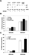
Knockdown of p21 and p27 restores the ability of FRNK-expressing endothelial cells to proliferate. HUVECs were electroporated with 150 to 200 ng of siRNA targeted toward luciferase (control), p21, and p27. The cells were then treated exactly as described in the legend to Fig. 9 and probed for p27 and p21 (A) and for the percentage of BrdU-positive cells (B). HUVECs were first electroporated with the specified siRNA as in panel A and on the following day during serum starvation were also transduced with Ad.GFP or Ad.GFP-FRNK. The next day, the cells were stimulated with complete growth medium for 16 h, at which point they were pulsed with BrdU and the percentage of BrdU-positive cells was calculated (C).

Similar articles

Cited by

References

    1. Abid, M. R., K. Yano, S. Guo, V. I. Patel, G. Shrikhande, K. C. Spokes, C. Ferran, and W. C. Aird. 2005. Forkhead transcription factors inhibit vascular smooth muscle cell proliferation and neointimal hyperplasia. J. Biol. Chem. 280:29864-29873. (First published 15 June 2005; doi:10.1074/jbc.M502149200.) - DOI - PubMed
    1. Abu-Ghazaleh, R., J. Kabir, H. Jia, M. Lobo, and I. Zachary. 2001. Src mediates stimulation by vascular endothelial growth factor of the phosphorylation of focal adhesion kinase at tyrosine 861, and migration and anti-apoptosis in endothelial cells. Biochem. J. 360:255-264. - PMC - PubMed
    1. Alavi, A., J. D. Hood, R. Frausto, D. G. Stupack, and D. A. Cheresh. 2003. Role of Raf in vascular protection from distinct apoptotic stimuli. Science 301:94-96. - PubMed
    1. Aplin, A. E., S. A. Stewart, R. K. Assoian, and R. L. Juliano. 2001. Integrin-mediated adhesion regulates ERK nuclear translocation and phosphorylation of Elk-1. J. Cell Biol. 153:273-282. - PMC - PubMed
    1. Assoian, R. K., and M. A. Schwartz. 2001. Coordinate signaling by integrins and receptor tyrosine kinases in the regulation of G1 phase cell-cycle progression. Curr. Opin. Genet. Dev. 11:48-53. - PubMed

Publication types

MeSH terms

Substances