. 2006 Jun;26(11):4214-25.
doi: 10.1128/MCB.01751-05.

Novel lymphotoxin alpha (LTalpha) knockout mice with unperturbed tumor necrosis factor expression: reassessing LTalpha biological functions

Affiliations

Novel lymphotoxin alpha (LTalpha) knockout mice with unperturbed tumor necrosis factor expression: reassessing LTalpha biological functions

Dmitry J Liepinsh et al. Mol Cell Biol. 2006 Jun.

Abstract

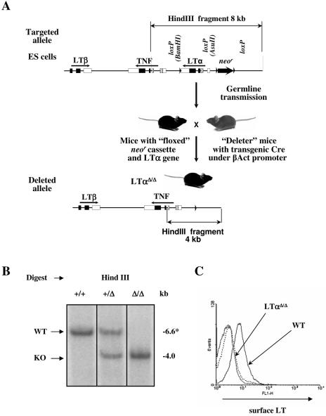
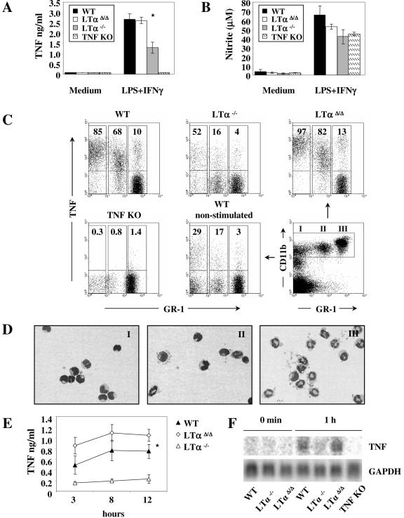
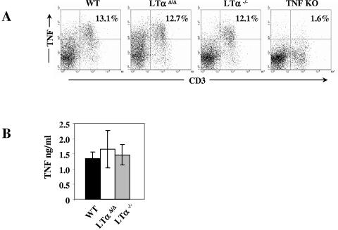
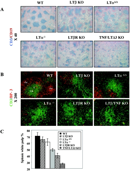
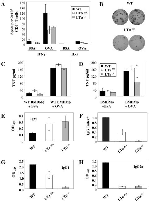
Lymphotoxin alpha (LTalpha) can exist in soluble form and exert tumor necrosis factor (TNF)-like activity through TNF receptors. Based on the phenotypes of knockout (KO) mice, the physiological functions of LTalpha and TNF are considered partly redundant, in particular, in supporting the microarchitecture of the spleen and in host defense. We exploited Cre-LoxP technology to generate a novel neomycin resistance gene (neo) cassette-free LTalpha-deficient mouse strain (neo-free LTalpha KO [LTalphaDelta/Delta]). Unlike the "conventional" LTalpha-/- mice, new LTalphaDelta/Delta animals were capable of producing normal levels of systemic TNF upon lipopolysaccharide (LPS) challenge and were susceptible to LPS/D-galactosamine (D-GalN) toxicity. Activated neutrophils, monocytes, and macrophages from LTalphaDelta/Delta mice expressed TNF normally at both the mRNA and protein levels as opposed to conventional LTalpha KO mice, which showed substantial decreases in TNF. Additionally, the spleens of the neo-free LTalpha KO mice displayed several features resembling those of LTbeta KO mice rather than conventional LTalpha KO animals. The phenotype of the new LTalphaDelta/Delta mice indicates that LTalpha plays a smaller role in lymphoid organ maintenance than previously thought and has no direct role in the regulation of TNF expression.


Figures

FIG. 1.
FIG. 1.
LoxP/Cre LTα gene targeting and deletion. (A) Scheme of the targeted disruption of the LTα gene. 129/Sv ES cells that have undergone homologous recombination with the targeting construct were injected into blastocysts and reimplanted into pseudopregnant females. The resulting chimeric mice were crossed with C57BL/6 mice to detect germ line transmission. After confirmation of germ line transmission by PCR and Southern analysis, the offspring were then crossed with transgenic “deleter” mice in which Cre recombinase was placed under the β-actin promoter to achieve deletion of both the neomycin resistance (neo) cassette and LTα gene. The progeny were tested for the LTα gene and neo deletion, intercrossed to obtain homozygous mice, and then backcrossed to the C57BL/6 background. (B) Southern blot analysis of splenocytes from LTα+/+, LTα+/Δ and LTαΔ/Δ mice. Genomic DNA was digested with HindIII. *, 6.6-kb fragment of wild-type mouse DNA with the floxed LTα gene. (C) Surface expression of the LTαβ complex on activated splenocytes. Splenocytes were plated at 0.5 × 10⁶/ml and activated for 7 h with phorbol myristate acetate (50 ng/ml) and anti-mouse CD40 (10 μg/ml).
FIG. 2.
FIG. 2.
Systemic LPS-induced TNF production is markedly decreased in conventional LTα−/−, but not in novel LTαΔ/Δ mice. Mice were injected intraperitoneally with 100 μg LPS. Serum TNF levels were measured by ELISA 90 min after injection. *, P < 0.001, wild-type versus LTα−/− mice; **, P < 0.001, LTαΔ/Δ versus LTα−/− mice.
FIG. 3.
FIG. 3.
Defective TNF synthesis by macrophages, monocytes, and neutrophils in the conventional LTα KO mice. (A) TNF and (B) nitric oxide (NO) production by BMDMφ. Cells were activated with either medium alone or LPS (10 ng/ml) and IFN-γ (10 ng/ml) for 24 h. *, P = 0.025, LTαΔ/Δ versus LTα−/−. (C) Intracellular TNF staining of whole blood activated with LPS (10 ng/ml) and IFN-γ (10 ng/ml) for 3 h. Events shown are gated for CD11bhi expression. (D) Wright-Giemsa-stained, sorted peripheral blood CD11bhi leukocytes subdivided into three separate populations according to GR-1 expression (representative untreated wild-type mouse). In population I, 90% of cells represent monocytes; population II consists of monocytes (50%), neutrophils (25%), and other cell types (25%); and population III is composed of neutrophils (96%). Percentages are average approximate values from multiple analyses. (E) TNF production by BMDNE. Cells were activated with 10 ng/ml LPS and IFN-γ. Supernatants were harvested at 3, 8, and 12 h. TNF was measured by sandwich ELISA. *, P = 0.019, wild-type versus LTα−/−. (F) Northern blot analysis of BMDNE stimulated with 10 ng/ml LPS and IFN-γ.
FIG. 4.
FIG. 4.
Normal TNF production by splenic T cells in both strains of LTα KO mice. (A) Intracellular TNF production by splenic T cells, and (B) soluble TNF production by purified T cells stimulated with phorbol myristate acetate (100 ng/ml) and ionomycin (1 μg/ml) for 5 h.
FIG. 5.
FIG. 5.
Better-conserved size of splenic white pulp and BP-3 expression in LTαΔ/Δ mice. (A) Serial splenic sections from mice of the indicated genotypes were stained with anti-CD3 (blue) and anti-CD19 (red) antibodies to detect T and B cells, respectively. Magnification, ×40. (B) To detect T cells and BP-3-positive stromal cells, splenic sections were stained with fluorescently labeled anti-CD3 (green) and anti-BP3 (red) antibodies. Magnification, ×200. (C) Quantification of the size of splenic white pulp. The size of splenic white pulp is shown as a percentage of the size of the entire image. Data were obtained using the NIH Medical Image Processing and Visualization program. Three mice per group were analyzed.
FIG. 6.
FIG. 6.
Adaptive immune responses of novel LTαΔ/Δ mice to OVA. (A) OVA- and BSA-specific IFN-γ and IL-5 production by sorted splenic CD4+ T cells from intraperitoneally OVA- and CFA-immunized mice. T cells were stimulated with OVA- or BSA-loaded fixed bone marrow-derived dendritic cells for 36 h. Antigen-specific cytokine production was determined by Elispot assay. (B) Representative wells showing OVA-specific IFN-γ production by splenic CD4+ T cells from wild-type and neo-free LTα KO mice. (C) TNF production by wild-type BMDMφ loaded with OVA or BSA (as a negative control) and stimulated for 24 h with purified day 14 CD4+ splenocytes from OVA/CFA-immunized wild-type (black columns), LTαΔ/Δ (white columns), or LTα−/− (gray columns) mice. (D) Antigen-specific TNF production by BMDMφ activated with wild-type CD4+ splenic T cells from OVA/CFA-immunized mice. *, P = 0.05, wild-type versus LTα−/−. (E to H) OVA-specific production of (E) IgM, (F) IgG, (G) IgG1, and (H) IgG2a was analyzed by ELISA. Serum dilution is 1:1,000. *, IgG response data representing three independent experiments. Readings at 405 nm were normalized to the wild-type reading, which was arbitrarily defined as 1.0 in each experiment.

Similar articles

Cited by

References

    1. Abe, K., F. O. Yarovinsky, T. Murakami, A. N. Shakhov, A. V. Tumanov, D. Ito, L. N. Drutskaya, K. Pfeffer, D. V. Kuprash, K. L. Komschlies, and S. A. Nedospasov. 2003. Distinct contributions of TNF and LT cytokines to the development of dendritic cells in vitro and their recruitment in vivo. Blood 101:1477-1483. - PubMed
    1. Aggarwal, B. B. 2003. Signalling pathways of the TNF superfamily: a double-edged sword. Nat. Rev. Immunol. 3:745-756. - PubMed
    1. Alexopoulou, L., M. Pasparakis, and G. Kollias. 1998. Complementation of lymphotoxin alpha knockout mice with tumor necrosis factor-expressing transgenes rectifies defective splenic structure and function. J. Exp. Med. 188:745-754. - PMC - PubMed
    1. Alimzhanov, M. B., D. V. Kuprash, M. H. Kosco-Vilbois, A. Luz, R. L. Turetskaya, A. Tarakhovsky, K. Rajewsky, S. A. Nedospasov, and K. Pfeffer. 1997. Abnormal development of secondary lymphoid tissues in lymphotoxin beta-deficient mice. Proc. Natl. Acad. Sci. USA 19;94:9302-9307. - PMC - PubMed
    1. Baer, M., A. Dillner, R. C. Schwartz, C. Sedon, S. A. Nedospasov, and P. F. Johnston. 1998. TNF alpha transcription in macrophages is attenuated by an autocrine factor that preferentially induces NF-κB p50. Mol. Cell. Biol. 18:5678-5689. - PMC - PubMed

Publication types

MeSH terms

LinkOut - more resources