. 2006 Jun;26(11):4316-26.
doi: 10.1128/MCB.02183-05.

Multisite protein kinase A and glycogen synthase kinase 3beta phosphorylation leads to Gli3 ubiquitination by SCFbetaTrCP

Affiliations

Multisite protein kinase A and glycogen synthase kinase 3beta phosphorylation leads to Gli3 ubiquitination by SCFbetaTrCP

Denis Tempé et al. Mol Cell Biol. 2006 Jun.

Abstract

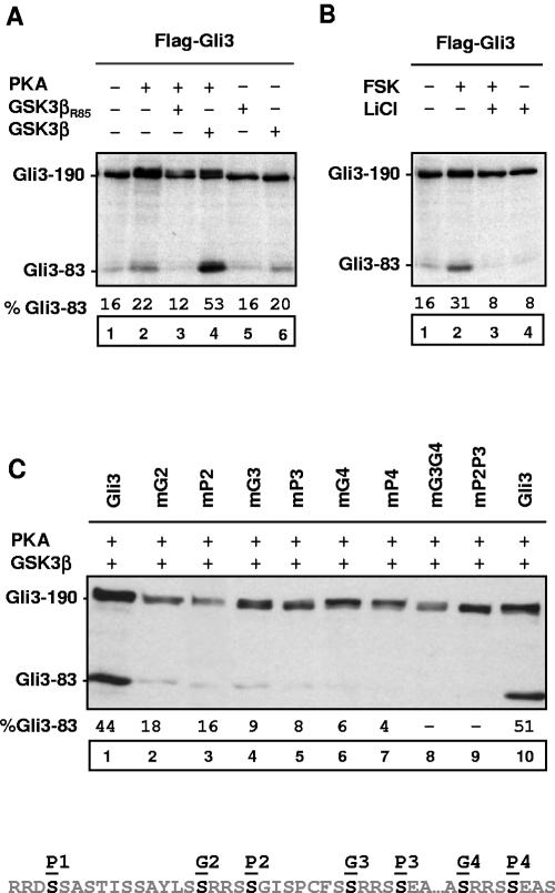
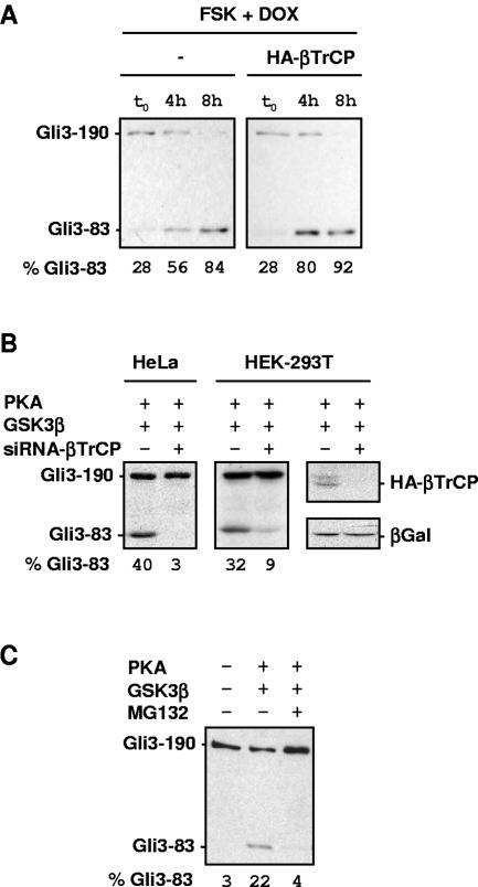
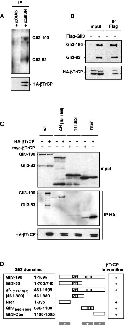
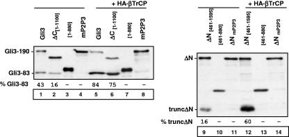
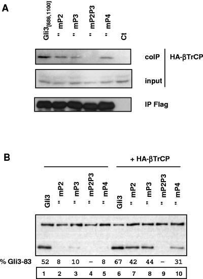
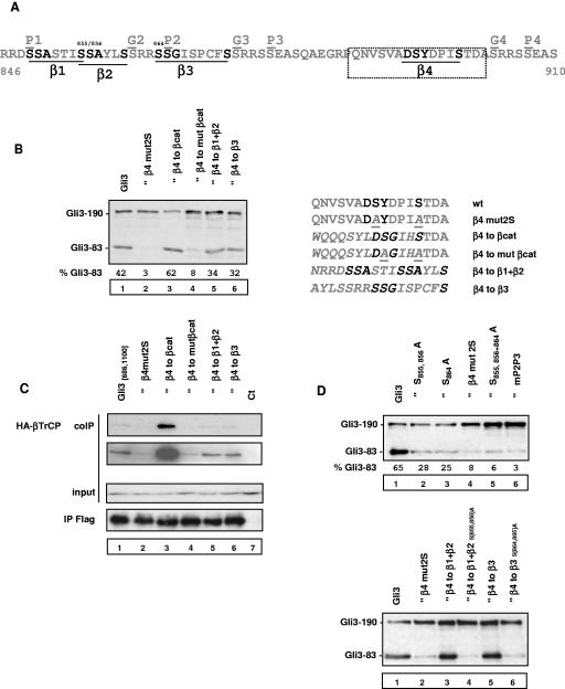
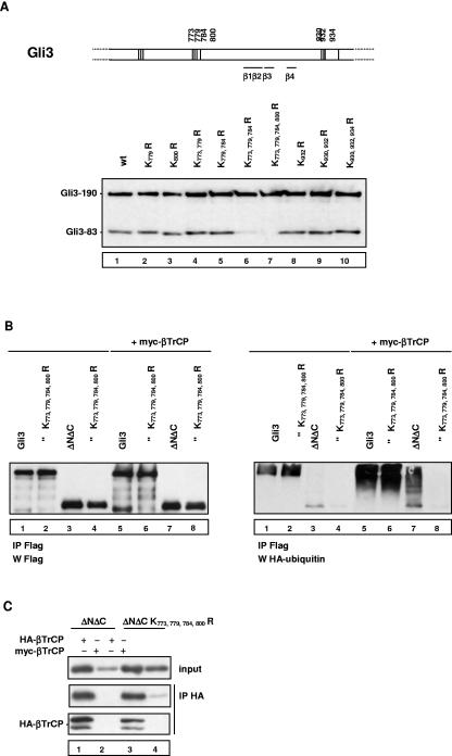
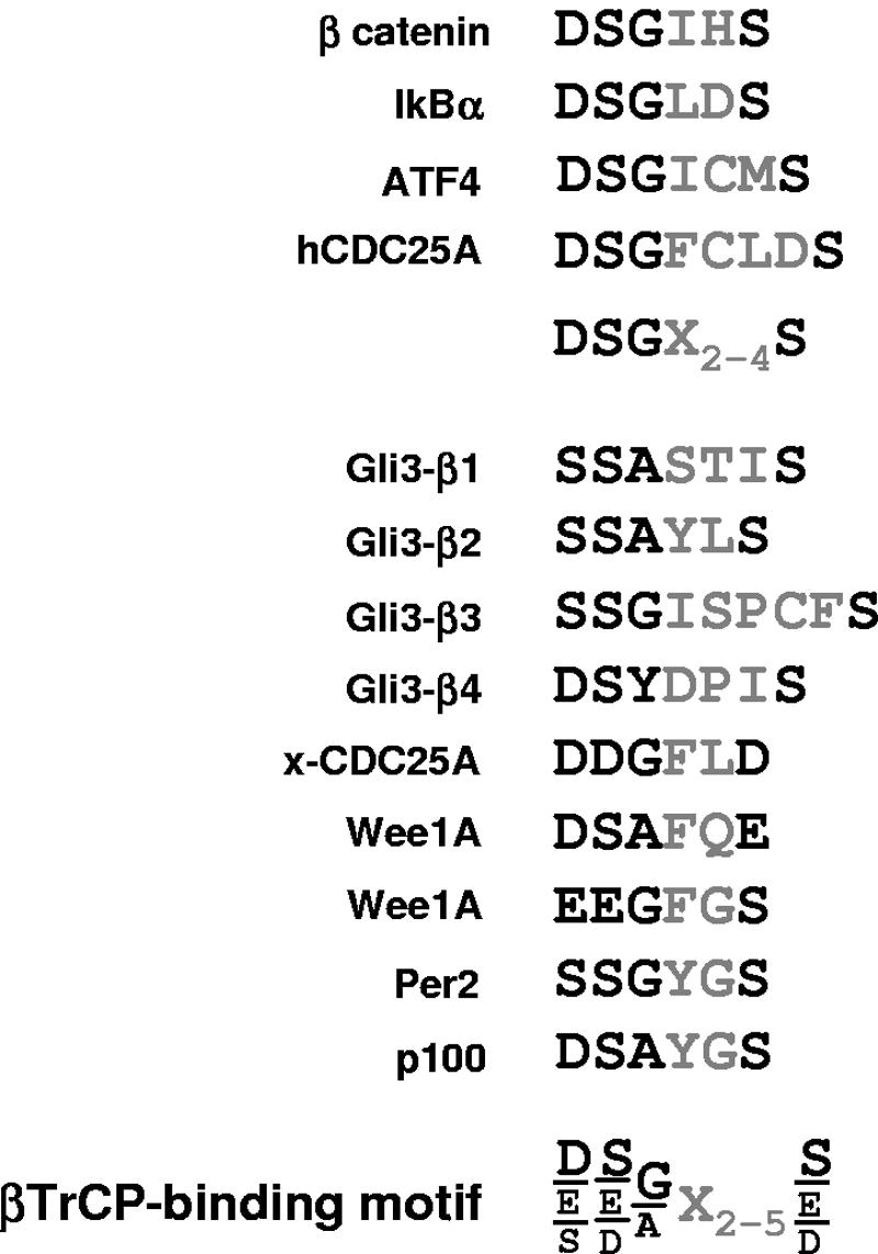
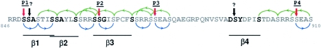
Gli3 is a zinc finger transcription factor proteolytically processed into a truncated repressor lacking C-terminal activation domains. Gli3 processing is stimulated by protein kinase A (PKA) and inhibited by Hedgehog signaling, a major signaling pathway in vertebrate development and disease. We show here that multisite glycogen synthase kinase 3beta (GSK3beta) phosphorylation and ubiquitination by SCFbetaTrCP are required for Gli3 processing. We identified multiple betaTrCP-binding sites related to the DSGX2-4S motif in Gli3, which are intertwined with PKA and GSK3beta sites, and SCFbetaTrCP target lysines that are essential for processing. Our results support a simple model whereby PKA triggers a cascade of Gli3 phosphorylation by GSK3beta and CK1 that leads to direct betaTrCP binding and ubiquitination by SCFbetaTrCP. Binding of betaTrCP to Gli3 N- and C-terminal domains lacking DSGX2-4S-related motifs was also observed, which could reflect indirect interaction via other components of Hedgehog signaling, such as the tumor suppressor Sufu. Gli3 therefore joins a small set of transcription factors whose processing is regulated by the ubiquitin-proteasome pathway. Our study sheds light on the role of PKA phosphorylation in Gli3 processing and will help to analyze how dose-dependent tuning of Gli3 processing is achieved by Hedgehog signaling.


Figures

FIG. 1.
FIG. 1.
Stimulation of truncated Gli3 synthesis by PKA and GSK3β. (A) Stimulation of truncated Gli3 synthesis by PKA and GSK3β stimulation. NIH 3T3 cells were transfected with expression vectors for Flag epitope-tagged Gli3 (Flag-Gli3), human PKA catalytic subunit (PKA), human wild-type GSK3β, or human dominant-negative GSK3β (GSK3βR85), and cell extracts were analyzed by immunoblotting with an anti-Flag antibody. (B) Inhibition of endogenous GSK3β by LiCl inhibits forskolin-induced synthesis of truncated Gli3. NIH 3T3 cells were transfected with expression vectors for Flag-Gli3. Where indicated, cells were treated either with 50 μM forskolin (FSK) alone or with 50 μM FSK and 20 mM LiCl for 12 h. Identical quantities of FSK vehicle (ethanol) were added to control cells. (C) Mutation of GSK3β sites adjacent to PKA sites inhibits synthesis of truncated Gli3. GSK3β phosphorylates serine or threonine residues that lie four residues N terminal to a phosphoserine. GSK3β sites SXXXPS, labeled G2 to G4, were found adjacent to PKA sites P2 to P4, respectively (29). Serine-to-alanine mutants at indicated sites of Flag-Gli3 were transfected into NIH 3T3 cells together with PKA and GSK3β expression vectors, and cell extracts were analyzed by immunoblotting with anti-Flag antibody. Autoradiograms were scanned to measure the signals corresponding to truncated and total Gli3 signals. The relative levels of truncated Gli3 are given as percentages of total (truncated + full length) Gli3 signal measured in each condition. A minus sign indicates lanes where truncated products were not quantified. Upon long exposure of blots, low-level processing could be detected and was roughly estimated to be inferior to 3% total Gli3. mG2, mP2, and mP2P3 indicate mutant Flag-Gli3 at site G2, site P2, and both sites P2 and P3, respectively.
FIG. 2.
FIG. 2.
Truncated Gli3 synthesis requires βTrCP. (A) Overexpression of wild-type βTrCP stimulates synthesis of truncated Gli3 in transcription shutoff experiments. NIH 3T3 cells were transfected with pBI-G-Flag-Gli3, pTet-Off, and GSK3β expression vectors together with control (−) or HA epitope-tagged βTrCP (HA-βTrCP) expression vectors. Cells were treated with 50 μM forskolin (FSK) and 100 ng/ml doxycycline (DOX) and collected after 0 h, 4 h, and 8 h to analyze the relative levels of truncated versus full-length Flag-Gli3. (B) Downregulation of βTrCP inhibits Gli3 processing. HeLa and 293T cells were transfected with siRNA against human βTrCP (+) or luciferase (−) together with Flag-Gli3, PKA, and GSK3β expression vectors. The right panel shows specific downregulation of HA-βTrCP by siRNA against βTrCP. 293T cells were transfected with siRNA against βTrCP (+) or luciferase (−) together with an expression vector for HA-βTrCP. Equal amounts of cell lysates were probed with anti-HA antibody to detect HA-βTrCP and anti-β-galactosidase (βGal) as a control of transfection and cytomegalovirus expression levels. (C) Gli3 processing is inhibited by MG132 proteasome inhibitor. NIH 3T3 cells were transfected with Flag-Gli3, PKAc, and GSK3β expression vectors. Cells were treated with 20 μM MG132 (+) or vehicle (−) for 6 h, and equal amounts of cell lysates were analyzed by anti-Flag immunoblotting.
FIG. 3.
FIG. 3.
Gli3 interacts with βTrCP. (A) Endogenous Gli3 interact with HA-βTrCP. C3H-10T1/2 cells were transfected with HA-βTrCP expression vector. The cell lysate from four plates of C3H-10T1/2 cells was split in half and subjected to parallel immunoprecipitations using control (αCtAb, control FGF4R antibody) or anti-Gli3N antibody. Immunoprecipitates were analyzed by immunoblotting with anti-HA antibody to detect HA-βTrCP or with anti-Gli3 antibody to detect endogenous Gli3. IP, immunoprecipitation. (B) HA-βTrCP coimmunoprecipitates with Flag-Gli3. NIH 3T3 cells were transfected with HA-βTrCP expression vector and control Flag or Flag-Gli3 expression vectors as indicated together with PKA and GSK3β expression vectors to stimulate synthesis of truncated Gli3. Equal amounts of cell lysates were subjected to anti-Flag immunoprecipitation and analyzed by immunoblotting. (C) Three different regions of Gli3 coimmunoprecipitate with HA-βTrCP. NIH 3T3 cells were transfected with expression vectors as indicated together with PKA and GSK3β expression vectors to stimulate synthesis of truncated Gli3. Plasmid mixtures contained either HA-βTrCP or βTrCP-myc expression constructs, the latter serving as negative controls in coimmunoprecipitation experiments. Cell lysates were subjected to βTrCP immunoprecipitation using anti-HA antibody followed by anti-Flag or anti-HA immunoblotting. In the bottom panel, the different constructs are represented. The position of PKA sites P1 to P6 essential for synthesis of truncated Gli3 (vertical bars) and the zinc finger region responsible for DNA binding (ZF) are indicated. βTrCP-binding sites inferred from analysis by coimmunoprecipitation with HA-βTrCP are indicated by plus signs. Experiments demonstrating that Gli3ΔN contains two independent binding sites to βTrCP are not shown (for Gli3 central 686-1100 domain binding to βTrCP, however, see Fig. 5). Gli3ΔN generated a truncated form upon stimulation by PKA and GSK3β that did not bind to βTrCP, most likely due to absence of the N-terminal βTrCP interaction domain. In the lane corresponding to Gli3 positions 461 to 880, we detected very low levels of truncated products, which could be due to low-level constitutive processing and which, accordingly, were not modulated by PKA, GSK3β, or βTrCP overexpression (data not shown). wt, wild type; Nter, N terminal; Cter, C terminal.
FIG. 4.
FIG. 4.
Gli3 N- and C-terminal domains are necessary for efficient processing. The indicated constructs were transfected into NIH 3T3 cells with PKA and GSK3β expression vectors, and the relative levels of truncated (trunc) and full-length products were analyzed by immunoblotting as described in the legend to Fig. 1. mP2P3 indicates serine-to-alanine mutations at PKA sites P2 and P3.
FIG. 5.
FIG. 5.
Effect of PKA site mutations on binding of βTrCP to Gli3 central 686-1100 domain. (A) Cells were transfected with HA-βTrCP, PKA, and GSK3β expression vectors and either wild-type Gli3 central 686-1100 domain, mutant Gli3 central 686-1100 domain, or control (ct) expression vectors as indicated. Cell lysates were subjected to anti-Flag immunoprecipitation (IP) and analyzed by immunoblotting. (B) The indicated constructs were transfected into NIH 3T3 cells with PKA and GSK3β expression vectors, and the relative levels of truncated and full-length products were analyzed by immunoblotting as described in the legend to Fig. 1. mG2, mP2, and mP2P3 indicate mutant Flag-Gli3 at site G2, site P2, and both sites P2 and P3, respectively.
FIG. 6.
FIG. 6.
Direct binding of βTrCP is required for Gli3 processing. (A) Identification of four sequence motifs related to the DSGX2-4S βTrCP-binding site in between PKA sites P1 and P4. SCFβTrCP substrates previously identified contain a DSGX2-4S sequence whose phosphorylation is necessary for βTrCP binding. The sequence motifs β1 to β4 underlined in the figure are related to the DSGX2-4S motif by alignment of the residues indicated in boldface. The 16-amino-acid sequence indicated by the box was mutated to test the role of motif β4 in processing and binding of βTrCP. (B) Effect of mutations in motif β4 on Gli3 processing. The indicated constructs were transfected into NIH 3T3 cells with PKA and GSK3β expression vectors, and the relative levels of truncated and full-length Gli3 were analyzed by immunoblotting. The constructs tested contained mutations of the 16-amino-acid box containing motif β4 as indicated in italics. The β-catenin (βcat) βTrCP-binding site was positioned such that the key serines are expected to be phosphorylated by sequential GSK3β activity after phosphorylation of site P4 by PKA (i.e., in a context mimicking their normal phosphorylation [1]). The mutant β-catenin motif does not bind βTrCP (15, 36). (C) Effect of mutations in motif β4 on binding of βTrCP to Gli3 central 686-1100 domain. NIH 3T3 cells were transfected with HA-βTrCP, PKA, and GSK3β expression vectors and wild-type (wt) or mutant (mut) Gli3 central 686-1100 domain expression vectors or control expression vector (ct) as indicated. Cell lysates were subjected to anti-Flag immunoprecipitation (IP) and analyzed by immunoblotting. Short and long exposures of immunoblots of coimmunoprecipitated (coIP) HA-βTrCP are shown. (D) Effect of mutations in motifs β1+β2 and β3 on Gli3 processing in their natural context (upper panel) or when replacing motif β4 (lower panel). The indicated constructs were transfected into NIH 3T3 cells with PKA and GSK3β expression vectors, and the relative levels of truncated and full-length Gli3 were analyzed by immunoblotting. mP2P3 indicates mutant Flag-Gli3 at PKA sites P2 and P3.
FIG. 7.
FIG. 7.
Identification of lysines essential for Gli3 processing that are ubiquitinated by SCFβTrCP. (A) Effect of single or multiple lysine-to-arginine mutations on Gli3 processing. The positions of lysines tested for their potential role in processing and ubiquitination are indicated. The position 846 to 910 domain containing βTrCP-binding motifs β1 to β4 does not contain any lysine. Lysines near this domain are represented by a vertical line. The indicated constructs were transfected into NIH 3T3 cells with PKA and GSK3β expression vectors, and the relative levels of truncated and full-length Gli3 were analyzed by immunoblotting. (B) Lysines 773, 779, 784, and 800 are essential for ubiquitination of Gli3ΔNΔC[461,1100] and its stimulation by βTrCP overexpression. The indicated Gli3 or Gli3ΔNΔC[461,1100] constructs were transfected into NIH 3T3 cells together with PKA, GSK3β, HA-ubiquitin, and control or myc-βTrCP expression vectors. Cells were treated with the proteasome inhibitor MG132 for 4 h at 20 μM, which inhibited proteolytic processing as described in Fig. 2C and favored detection of ubiquitinated proteins, and cells were lysed in lysis buffer containing 5% SDS at 95°C for 10 min. The resulting extracts were subjected to immunoprecipitation with anti-Flag antibody and analyzed by immunoblotting with anti-HA antibody to detect ubiquitinated species (right panel) or with anti-Flag antibody (left panel, labeled “IP Flag, W Flag”). (C) Mutation of lysines 773, 779, 784, and 800 to arginine does not impair binding of Gli3ΔNΔC[461,1100] to HA-βTrCP. NIH 3T3 cells were transfected with expression vectors as indicated together with PKA and GSK3β expression vectors. Cell lysates were subjected to βTrCP immunoprecipitation (IP) using anti-HA antibody followed by anti-Flag or anti-HA immunoblotting. wt, wild type.
FIG. 8.
FIG. 8.
Alignment of βTrCP-binding motifs in known SCFβTrCP substrates. The first SCFβTrCP substrates identified allowed defining a common DSGX2-4S sequence whose phosphorylation is necessary for βTrCP binding (11). Alignment of βTrCP-binding sites in recently identified substrates allows proposing a revised consensus βTrCP-binding motif. For hCDC25A, Per2, p100, xCDC25A, and Wee1A substrates, see references , , , , and , respectively.
FIG. 9.
FIG. 9.
Phosphorylation of βTrCP-binding sites by a putative cascade of GSK3β, CK1, and PKA phosphorylations. PKA phosphorylates serines in consensus RRXS sites indicated by red arrows. GSK3β phosphorylates serines four residues N terminal to a phosphoserine, while CK1 phosphorylates serines three residues C terminal to a phosphoserine; both can sequentially multiphosphorylate substrates after priming (1, 9, 14). S855 in motif β1 could, therefore, be phosphorylated as follows: S849 (P1) phosphorylation by PKA priming sequential phosphorylation of S852 and S855 by CK1. S856 phosphorylation in motif β2 could be as follows: S865 (P2) by PKA priming S868 by CK1 and then S864, S860, and S856 by GSK3β. Similar phosphorylation pathways can easily be proposed for all serines in β1 to β4 motifs (blue and green arrows representing phosphorylations by CK1 and GSK3β, respectively), except S850 and S894 (indicated by black arrows). S850 and S894 lack serines at n + 4 or n − 3 positions for phosphorylation priming, and their sequence context is not similar to that in unprimed CK1 sites (14). An alternative candidate kinase is Fused. S850 phosphorylation may not be required if βTrCP could bind to the overlapping DSS850ASTIS motif (with S850 aligned to G/A in the consensus) rather than the motif proposed in Fig. 8. In any case, it appears that 19 serines in the 65-amino-acid segment from P1 to P4, including most serines in βTrCP-binding motifs β1 to β4, are likely phosphorylated by GSK3β and CK1 after priming by PKA.

Similar articles

Cited by

References

    1. Amit, S., A. Hatzubai, Y. Birman, J. S. Andersen, E. Ben-Shushan, M. Mann, Y. Ben-Neriah, and I. Alkalay. 2002. Axin-mediated CKI phosphorylation of beta-catenin at Ser 45: a molecular switch for the Wnt pathway. Genes Dev. 16:1066-1076. - PMC - PubMed
    1. Bai, C. B., D. Stephen, and A. L. Joyner. 2004. All mouse ventral spinal cord patterning by hedgehog is Gli dependent and involves an activator function of Gli3. Dev. Cell 6:103-115. - PubMed
    1. Busino, L., M. Donzelli, M. Chiesa, D. Guardavaccaro, D. Ganoth, N. V. Dorrello, A. Hershko, M. Pagano, and G. F. Draetta. 2003. Degradation of Cdc25A by beta-TrCP during S phase and in response to DNA damage. Nature 426:87-91. - PubMed
    1. Chen, C. H., D. P. von Kessler, W. Park, B. Wang, Y. Ma, and P. A. Beachy. 1999. Nuclear trafficking of Cubitus interruptus in the transcriptional regulation of Hedgehog target gene expression. Cell 98:305-316. - PubMed
    1. Chen, Y., N. Gallaher, R. H. Goodman, and S. M. Smolik. 1998. Protein kinase A directly regulates the activity and proteolysis of cubitus interruptus. Proc. Natl. Acad. Sci. USA 95:2349-2354. - PMC - PubMed

Publication types

MeSH terms