

A theoretical analysis of water transport through chondrocytes
- PMID: 16705444
- PMCID: PMC2853978
- DOI: 10.1007/s10237-006-0039-9
A theoretical analysis of water transport through chondrocytes
Abstract
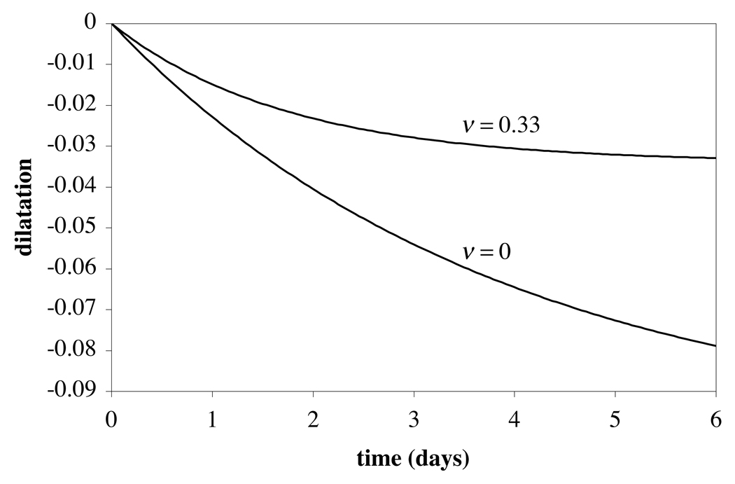
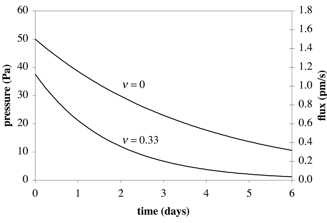
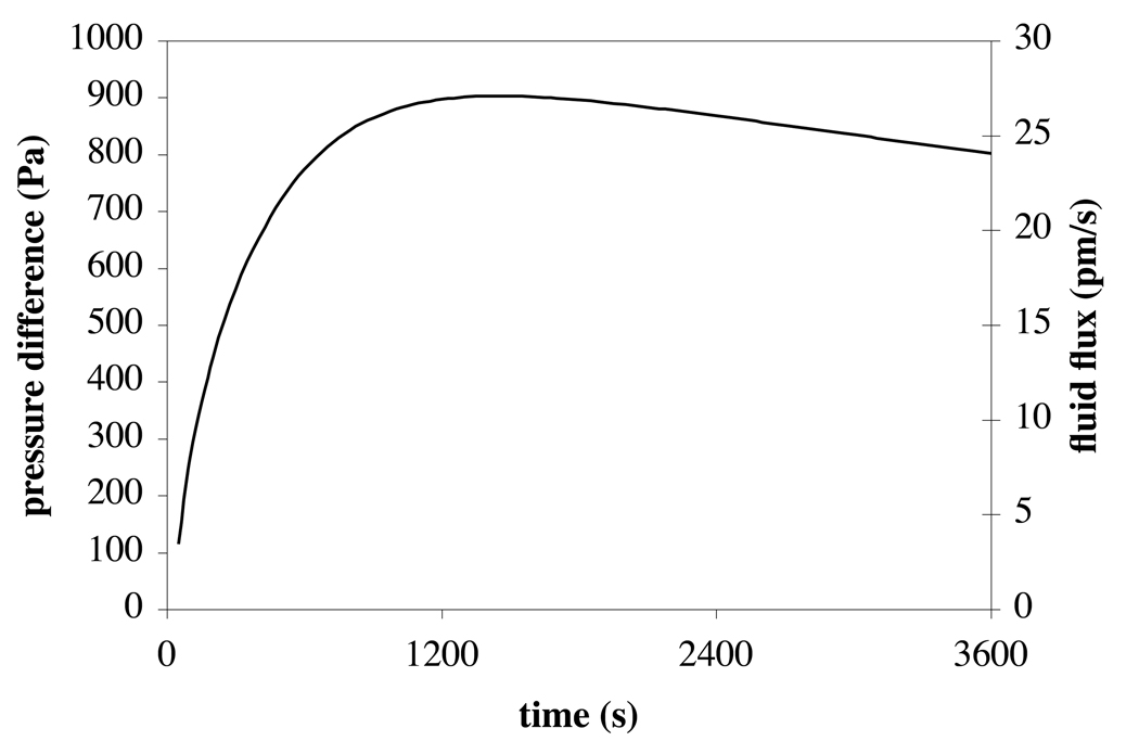
Because of the avascular nature of adult cartilage, nutrients and waste products are transported to and from the chondrocytes by diffusion and convection through the extracellular matrix. The convective interstitial fluid flow within and around chondrocytes is poorly understood. This theoretical study demonstrates that the incorporation of a semi-permeable membrane when modeling the chondrocyte leads to the following findings: under mechanical loading of an isolated chondrocyte the intracellular fluid pressure is on the order of tens of Pascals and the transmembrane fluid outflow, on the order of picometers per second, takes several days to subside; consequently, the chondrocyte behaves practically as an incompressible solid whenever the loading duration is on the order of minutes or hours. When embedded in its extracellular matrix (ECM), the chondrocyte response is substantially different. Mechanical loading of the tissue leads to a fluid pressure difference between intracellular and extracellular compartments on the order of tens of kilopascals and the transmembrane outflow, on the order of a nanometer per second, subsides in about 1 h. The volume of the chondrocyte decreases concomitantly with that of the ECM. The interstitial fluid flow in the extracellular matrix is directed around the cell, with peak values on the order of tens of nanometers per second. The viscous fluid shear stress acting on the cell surface is several orders of magnitude smaller than the solid matrix shear stresses resulting from the ECM deformation. These results provide new insight toward our understanding of water transport in chondrocytes.
Figures


Similar articles
-
Shape of chondrocytes within articular cartilage affects the solid but not the fluid microenvironment under unconfined compression.Acta Biomater. 2016 Jan;29:170-179. doi: 10.1016/j.actbio.2015.10.035. Epub 2015 Oct 23. Acta Biomater. 2016. PMID: 26525115 Free PMC article.
-
The effect of matrix tension-compression nonlinearity and fixed negative charges on chondrocyte responses in cartilage.Mol Cell Biomech. 2005 Dec;2(4):191-204. Mol Cell Biomech. 2005. PMID: 16705865
-
The mechanical environment of the chondrocyte: a biphasic finite element model of cell-matrix interactions in articular cartilage.J Biomech. 2000 Dec;33(12):1663-73. J Biomech. 2000. PMID: 11006391
-
Effects of shear stress on articular chondrocyte metabolism.Biorheology. 2000;37(1-2):95-107. Biorheology. 2000. PMID: 10912182 Review.
-
The deformation behavior and viscoelastic properties of chondrocytes in articular cartilage.Biorheology. 2000;37(1-2):27-44. Biorheology. 2000. PMID: 10912176 Review.
Cited by
-
Shear stress induced by fluid flow produces improvements in tissue-engineered cartilage.Biofabrication. 2020 Aug 10;12(4):045010. doi: 10.1088/1758-5090/aba412. Biofabrication. 2020. PMID: 32640430 Free PMC article.
-
Primary cilium-mediated mechanotransduction in cartilage chondrocytes.Exp Biol Med (Maywood). 2023 Aug;248(15):1279-1287. doi: 10.1177/15353702231199079. Epub 2023 Oct 28. Exp Biol Med (Maywood). 2023. PMID: 37897221 Free PMC article. Review.
-
Computational modeling of chemical reactions and interstitial growth and remodeling involving charged solutes and solid-bound molecules.Biomech Model Mechanobiol. 2014 Oct;13(5):1105-20. doi: 10.1007/s10237-014-0560-1. Epub 2014 Feb 21. Biomech Model Mechanobiol. 2014. PMID: 24558059 Free PMC article.
-
Chondroitin Sulfate-Tyramine-Based Hydrogels for Cartilage Tissue Repair.Int J Mol Sci. 2023 Feb 9;24(4):3451. doi: 10.3390/ijms24043451. Int J Mol Sci. 2023. PMID: 36834862 Free PMC article.
-
Mechanical Loading of Cartilage Explants with Compression and Sliding Motion Modulates Gene Expression of Lubricin and Catabolic Enzymes.Cartilage. 2015 Jul;6(3):185-93. doi: 10.1177/1947603515581680. Cartilage. 2015. PMID: 26175864 Free PMC article.
References
-
- Almeida ES, Spilker RL. Mixed and Penalty Finite Element Models for the Nonlinear Behavior of Biphasic Soft Tissues in Finite Deformation: Part I - Alternate Formulations. Comput Methods Biomech Biomed Engin. 1997;1:25–46. - PubMed
-
- Andarawis NA, Seyhan SL, Mauck RL, Soltz MA, Ateshian GA, Hung CT. A novel permeation device for hydrogels and soft tissues; Proceedings of the 2001 ASME International Mechanical Engineering Congress and Exposition; 2001. p. 23149.
-
- Armstrong CG, Lai WM, Mow VC. An analysis of the unconfined compression of articular cartilage. J Biomech Eng. 1984;106:165–173. - PubMed
-
- Ateshian GA, Lai WM, Zhu WB, Mow VC. An asymptotic solution for the contact of two biphasic cartilage layers. J Biomech. 1994;27:1347–1360. - PubMed
-
- Ateshian GA, Wang H. A theoretical solution for the frictionless rolling contact of cylindrical biphasic articular cartilage layers. J Biomech. 1995;28:1341–1355. - PubMed
Publication types
MeSH terms
Substances
Grants and funding
LinkOut - more resources
Full Text Sources
Other Literature Sources