

Diesel exhaust particles induce the over expression of tumor necrosis factor-alpha (TNF-alpha) gene in alveolar macrophages and failed to induce apoptosis through activation of nuclear factor-kappaB (NF-kappaB)
- PMID: 16705808
- PMCID: PMC3814704
- DOI: 10.3390/ijerph2005010107
Diesel exhaust particles induce the over expression of tumor necrosis factor-alpha (TNF-alpha) gene in alveolar macrophages and failed to induce apoptosis through activation of nuclear factor-kappaB (NF-kappaB)
Abstract
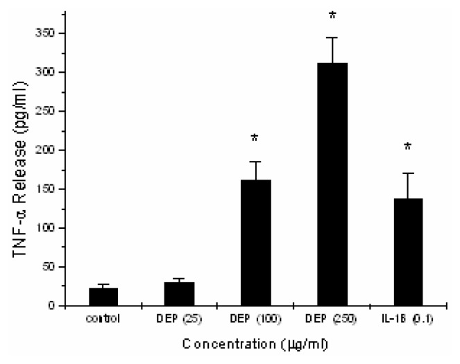
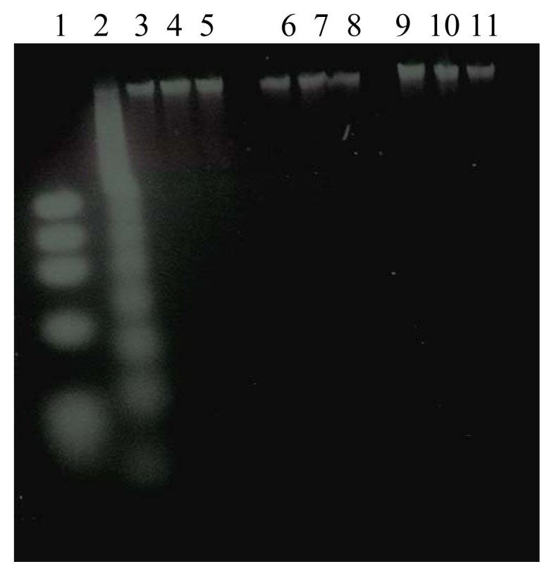
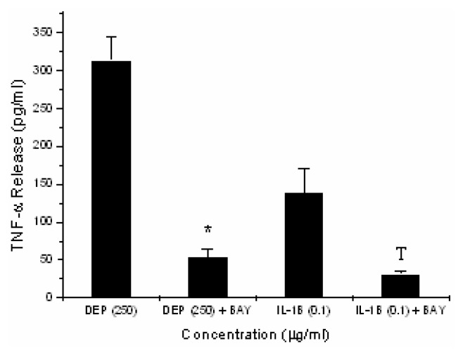
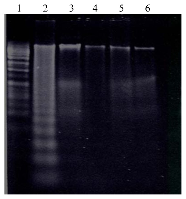
Exposure to particulate matter (PM2.5-10), including diesel exhaust particles (DEP) has been reported to induce lung injury and exacerbation of asthma and chronic obstructive pulmonary disease. Alveolar macrophages play a major role in the lung's response to inhaled particles and therefore, are a primary target for PM2.5-10 effect. The molecular and cellular events underlying DEP-induced toxicity in the lung, however, remain unclear. To determine the effect of DEP on alveolar macrophages, RAW 264.7 cells were grown in RPMI 1640 with supplements until confluency. RAW 264.7 cultures were exposed to Hank's buffered saline solution (vehicle), vehicle containing an NF-kappaB inhibitor, BAY11-7082 (25 microM with 11/2 hr pre-incubation), or vehicle containing DEP (250 microg/ml) in the presence or absence of BAY11-7082 (25 microM with 11/2 hr pre-incubation) for 4 hr and TNF-alpha release was determined by enzyme-linked immunosorbent assay and confirmed by western blots. RAW 264.7 apoptotic response was determined by DNA fragmentation assays. U937 cells treated with campothecin (4 microg/ml x 3 hr), an apoptosis-inducing agent, were used as positive control. We report that exposure to the carbonaceous core of DEP induces significant release of TNF-alpha in a concentration-dependent fashion (31 +/- 4 pg/ml, n = 4, p = 0.08; 162 +/- 23 pg/ml, n = 4, p < 0.05; 313 +/- 31 pg/ml, n = 4, p < 0.05 at 25, 100, and 250 microg/ml, respectively). DEP exposure, however, failed to induce any apoptotic response in RAW 264.7 cells. Moreover, inhibition of NF-kappaB binding activity has resulted in DEP-induced apoptotic response in alveolar macrophages, as demonstrated by the NF-kappaB inhibitor, BAY11-7082 studies. The results of the present study indicate that DEP induce the release of TNF-alpha in alveolar macrophages, a primary target for inhaled particles effect. DEP-induced TNF-alpha gene expression is regulated at the transcriptional level by NF-kappaB. Furthermore, DEP-induced increase in NF-kappaB-DNA binding activity appears to protect against apoptosis.
Figures

References
-
- Ohtoshi T., Tazikawa H., Okazaki H., Kawasaki S., Takeuchi N., Ohta K., Ito K. Diesel exhaust particles (DEP) stimulate human airway epithelial cells to produce cytokines relevant to airway inflammation in vitro. J. Allergy Clin. Immunol. 1998;101:778–785. - PubMed
-
- Sagai M., Furuyama A., Ichinose T. Biological effects of diesel exhaust particles (DEP) HI. Pathogenesis of asthma-like symptoms in mice. Free Rod. Biol. Med. 1996;21:199. - PubMed
-
- Nel A. E., Diaz-Sanchez D., Ng D., Hiura J., Saxon A. Enhancement of allergic inflammation by the interaction between diesel exhaust particles and the immune system. J. Allergy Clin. Immunol. 1998;102:539–554. - PubMed
-
- Peterson B. A., Saxon A. Global increases in allergic respiratory disease: The possible roles of diesel exhaust particles. Ann. Allergy Asthma Immunol. 1996;77(4):263–268. - PubMed
-
- Diaz-Sanchez D. The roles of diesel exhaust particles and their associated polyaromatic hydrocarbons in the induction of allergic airway disease. Allergy. 1997;52(38s):52–56. - PubMed
Publication types
MeSH terms
Substances
Grants and funding
LinkOut - more resources
Full Text Sources
Other Literature Sources