

Cytokines and adhesion molecules expression in the brain in human cerebral malaria
- PMID: 16705810
- PMCID: PMC3814706
- DOI: 10.3390/ijerph2005010123
Cytokines and adhesion molecules expression in the brain in human cerebral malaria
Abstract
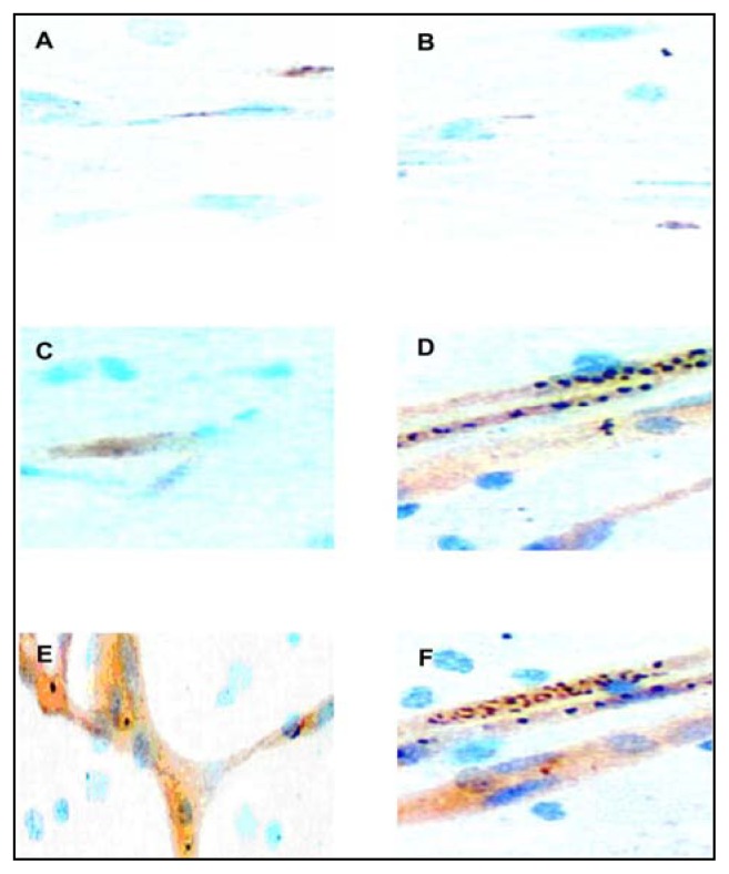
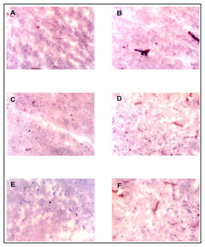
Although the role of systemic proinflammatory cytokines, IL-1beta and TNF-alpha, and their up-regulation of adhesion molecules, ICAM-1, VCAM-1 and E-Selectin, in the pathogenesis of cerebral malaria (CM) is well established, the role of local cytokine release remain unclear. Immunohistochemistry (IHC) was used to compare the expression of ICAM-1, VCAM-1, E-Selectin, IL-1beta, TNF-a and TGF-beta at light microscopic level in cerebral, cerebellar and brainstem postmortem cryostat sections from 10 CM, 5 severe malarial anemia (SMA), 1 purulent bacterial meningitis (PBM), 2 non-central nervous system infections (NCNSI) and 3 non-infections (NI) deaths in Ghanaian children. Fatal malaria and Salmonella sepsis showed significantly higher vascular expression of all 3 adhesion molecules, with highly significant co-localization with sequestration in the malaria cases. However, there was negligible difference between CM and SMA. TGF-beta showed intravascular and perivascular distribution in all cases, but expression was most intense in the PBM case and CM group. TNF-alpha and IL-1beta showed prominent brain parenchymal staining, in addition to intravascular and perivascular staining, in only the PBM case and CM group. The maximal expression of all 6 antigens studied was in the cerebellar sections of the malaria cases. Endothelial activation is a feature of fatal malaria and Salmonella sepsis, with adhesion molecule expression being highly correlated with sequestration. IL-1beta and TNF-alpha are upregulated in only cases with neurodegenerative lesions, whilst TGF-beta is present in all cases. Both cytokines and adhesion molecules were maximally upregulated in the cerebellar sections of the malaria cases.
Figures
References
-
- Tropical Disease Research (TDR) Tropical Research. 7th Program Report, UNDPA World Bank/WHO; Geneva: 1998. Malaria; pp. 3–40.
-
- Kwiatkowski D., Hill A. V., Sambou I., Twumasi P., Castracane J., Manogue K. R., Cerami A., Brewster D. R., Greenwood B. M. TNF concentration in fatal cerebral, non-fatal cerebral and uncomplicated Plasmodium falciparum malaria. Lancet. 1990;336:1201–1204. - PubMed
-
- Newton C. R. J. C., Krishna S. Severe falciparum malaria in children: current understanding of pathophysiology and supportive treatment. Pharmacol Ther. 1998;79:1–53. - PubMed
-
- Brown H., Turner G., Rogerson S., Tembo M., Mwenechanya J., Molyneux M., Taylor T. Cytokine Expression in the Brain in Human Cerebral Malaria. J Infect Dis. 1999;180:1742–1746. - PubMed
MeSH terms
Substances
LinkOut - more resources
Full Text Sources
Miscellaneous