. 2006 May 30;103(22):8499-504.
doi: 10.1073/pnas.0602957103. Epub 2006 May 17.

Inhibition of dsRNA-induced signaling in hepatitis C virus-infected cells by NS3 protease-dependent and -independent mechanisms

Affiliations

Inhibition of dsRNA-induced signaling in hepatitis C virus-infected cells by NS3 protease-dependent and -independent mechanisms

Guofeng Cheng et al. Proc Natl Acad Sci U S A. .

Abstract

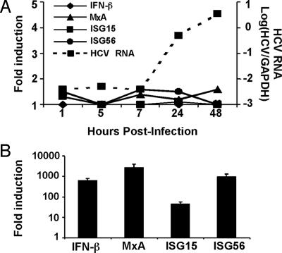
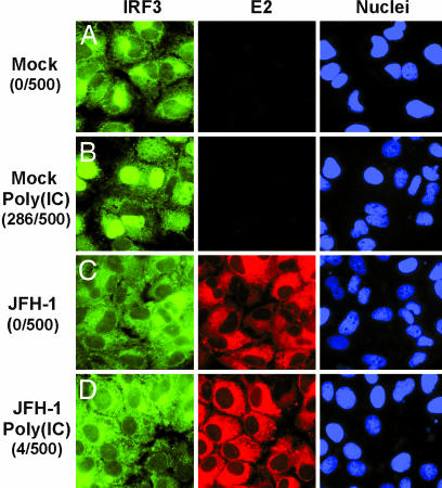
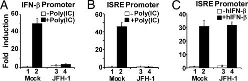
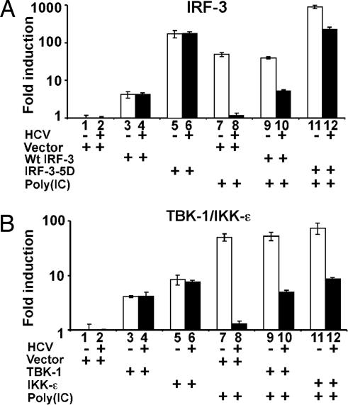
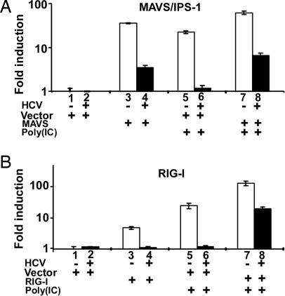
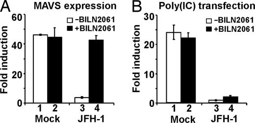
The recent establishment of a robust hepatitis C virus (HCV) cell culture system permits analysis of virus-host interactions during HCV infection. Here, we report that HCV genotype 2a (JFH-1) infection fails to induce IFN-beta or IFN-stimulated gene expression in Huh-7 cells, and that it blocks IFN-beta and IFN-stimulated gene production after transfection of synthetic dsRNA. Overexpression of individual components of the dsRNA-signaling pathway in HCV-infected and uninfected cells indicates that HCV inhibits IFN-beta promoter activity by inactivating the mitochondrial antiviral signaling protein/IFN-beta promoter stimulator 1 (MAVS/IPS-1), while leaving the IFN-induced Janus kinases-signal transducers and activators of transcription (JAK-STAT) signaling pathway intact. We also show that MAVS/IPS-1-dependent IFN-beta promoter activity in HCV-infected cells is fully restored by the nonstructural protein 3 (NS3) protease inhibitor BILN2061. In contrast, synthetic dsRNA-induced IFN-beta promoter activity is not restored by BILN2061, although it is partially restored by overexpression of RIG-I. These results support recently reported evidence that the HCV NS3 protease blunts the ability of HCV to induce IFN-beta promoter activity by proteolytically cleaving MAVS/IPS-1. The results also suggest that HCV blocks the synthetic dsRNA-induced signaling pathway at a point upstream of MAVS/IPS-1, and that it does so by an NS3-independent mechanism.


Conflict of interest statement

Conflict of interest statement: F.V.C. consults for several companies that are developing antiviral drugs and/or vaccines for HCV.

Figures

Fig. 1.
Fig. 1.
HCV infection does not induce IFN-β or ISG expression. (A) RT-qPCR analysis of cellular HCV RNA, IFN-β, and ISG expression. Huh-7 cells were infected with JFH-1 at a high multiplicity (moi = 3). At the indicated times, total cellular RNA was harvested and extracted. The intracellular gene expression of IFN-β, ISGs (MxA, ISG15 and ISG56), and HCV viral RNA was then measured by RT-qPCR. The expression of IFN-β or ISGs is presented as fold-induction relative to basal levels in uninfected cells. (B) Induction of ISGs in Huh-7 cells after transfection with poly(IC). Huh-7 cells were transfected with poly(IC) by using Lipofectamine 2000. Sixteen hours later, total cellular RNA was harvested. Quantification of the intracellular gene expression of ISGs was performed the same as in A.
Fig. 2.
Fig. 2.
HCV infection prevents dsRNA-induced IRF-3 nuclear translocation. Huh-7 cells were either mock-infected (A and B) or infected with JFH-1 at moi = 0.2 (C and D). On day 4 postinfection, cells were either transfected with poly(IC) by using Lipofectamine 2000 (B and D) or left untransfected (A and C). Sixteen hours later, cells were fixed and stained with antibodies against IRF-3 (green) and HCV E2 (red). Cell nuclei were stained by Hoechst dye (blue). To measure the nuclear accumulation of IRF-3, 500 cells from ten independent fields were examined on a fluorescent microscope, and the results are shown on the left in parentheses as the number of cells with nuclear IRF-3 per 500 cells.
Fig. 3.
Fig. 3.
Blockade of dsRNA-induced IFN-β expression in HCV-infected cells. (A) HCV blocks dsRNA-induced IFN-β promoter activation. Huh-7 cells were infected with JFH-1 at moi = 0.2. On day 4 postinfection, cells were cotransfected with the IFN-β promoter luciferase reporter plasmid, an internal control plasmid pRL-TK, and a carrier plasmid pcDNA3.1. Thirty-six hours later, cells were transfected with or without poly(IC). Cells were subjected to dual luciferase assay 16 h after transfection. The results are expressed as fold induction of IFN-β promoter activity relative to the basal level. (B) HCV blocks dsRNA-induced ISRE promoter activation. The experiment was performed the same as in A, except that the ISRE promoter luciferase reporter plasmid was used. (C) HCV does not block IFN-induced JAK-STAT signaling. Similar to B, cells were transfected with the ISRE promoter reporter plasmid and then incubated with 100 units per ml of human IFN-β for 16 h before dual luciferase assay.
Fig. 4.
Fig. 4.
HCV blocks the dsRNA-signaling pathway upstream of the IRF-3 kinases TBK-1/IKK-ε. Huh-7 cells were infected with JFH-1 at moi = 0.2. On day 4 postinfection, cells were cotransfected with the reporter plasmid pIFΔ(−125) lucter and plasmid pRL-TK in the presence of an empty vector, or a plasmid expressing wild-type IRF-3 or its active mutant IRF-3–5D (A), or with plasmids expressing the TBK-1 or IKK-ε (B) as indicated. Thirty-six hours later, cells were transfected with or without poly(IC). Luciferase activities were measured 16 h after transfection, and the results are expressed as fold induction of IFN-β promoter activity.
Fig. 5.
Fig. 5.
HCV blocks MAVS/IPS-1- and RIG-I-induced IFN-β expression. HCV blocks MAVS/IPS-1-mediated (A) or RIG-I-mediated (B) IFN-β induction. The experimental conditions were the same as in Fig. 4, except that MAVS/IPS-1 or RIG-I expression plasmids were cotransfected with the IFN-β promoter reporter plasmid.
Fig. 6.
Fig. 6.
The NS3 protease inhibitor BILN2061 restores MAVS/IPS-1-mediated, but not dsRNA-mediated, IFN-β induction in cells infected with HCV. (A) NS3 protease inhibitor restores the MAVS/IPS-1-mediated IFN-β induction. Twenty-four hours before cotransfection with the MAVS/IPS-1 expression vector and the IFN-β promoter reporter plasmid, the NS3 protease inhibitor BILN2061 was added to mock infected and JFH-1 infected cells at a final concentration of 10 μM. Luciferase activities were analyzed 36 h after transfection in the absence of poly(IC). (B) NS3 protease inhibitor fails to restore the dsRNA-mediated IFN-β induction. Similar to A, cells were pretreated with the protease inhibitor BILN2061 before plasmid transfection. Cells were further transfected with or without poly(IC) 36 h after transfection of the IFN-β promoter reporter plasmid. Sixteen hours later, luciferase activities were analyzed, and the results are expressed as fold induction.

References

    1. Hoofnagle J. H. Hepatology. 2002;36:S21–S29. - PubMed
    1. Alter H. J., Seeff L. B. Semin. Liver Dis. 2000;20:17–35. - PubMed
    1. Zhong J., Gastaminza P., Cheng G., Kapadia S., Kato T., Burton D. R., Wieland S. F., Uprichard S. L., Wakita T., Chisari F. V. Proc. Natl. Acad. Sci. USA. 2005;102:9294–9499. - PMC - PubMed
    1. Lindenbach B. D., Evans M. J., Syder A. J., Wolk B., Tellinghuisen T. L., Liu C. C., Maruyama T., Hynes R. O., Burton D. R., McKeating J. A., Rice C. M. Science. 2005;309:623–626. - PubMed
    1. Wakita T., Pietschmann T., Kato T., Date T., Miyamoto M., Zhao Z., Murthy K., Habermann A., Krausslich H. G., Mizokami M., et al. Nat. Med. 2005;11:791–796. - PMC - PubMed

Publication types

MeSH terms

LinkOut - more resources