. 2006 May 30;103(22):8342-7.
doi: 10.1073/pnas.0602598103. Epub 2006 May 17.

Locating active-site hydrogen atoms in D-xylose isomerase: time-of-flight neutron diffraction

Affiliations

Locating active-site hydrogen atoms in D-xylose isomerase: time-of-flight neutron diffraction

Amy K Katz et al. Proc Natl Acad Sci U S A. .

Abstract

Time-of-flight neutron diffraction has been used to locate hydrogen atoms that define the ionization states of amino acids in crystals of D-xylose isomerase. This enzyme, from Streptomyces rubiginosus, is one of the largest enzymes studied to date at high resolution (1.8 A) by this method. We have determined the position and orientation of a metal ion-bound water molecule that is located in the active site of the enzyme; this water has been thought to be involved in the isomerization step in which D-xylose is converted to D-xylulose or D-glucose to D-fructose. It is shown to be water (rather than a hydroxyl group) under the conditions of measurement (pH 8.0). Our analyses also reveal that one lysine probably has an -NH(2)-terminal group (rather than NH(3)(+)). The ionization state of each histidine residue also was determined. High-resolution x-ray studies (at 0.94 A) indicate disorder in some side chains when a truncated substrate is bound and suggest how some side chains might move during catalysis. This combination of time-of-flight neutron diffraction and x-ray diffraction can contribute greatly to the elucidation of enzyme mechanisms.


Conflict of interest statement

Conflict of interest statement: No conflicts declared.

Figures
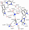
Fig. 1.
Fig. 1.
Active site of XI showing most of the amino acid residues described in this work. The two Trp and two His residues that define the “box” are drawn with black-filled bonds, as is Glu-217, which is shared by both metal ions, M1 and M2.
Fig. 2.
Fig. 2.
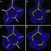
Doubly protonated His residues in 2FoFc nuclear-density maps (1.8-Å resolution) (a and c) and 2FoFc electron-density maps (0.94 Å resolution) (b and d) for His-54 (a and b) and His-220 (c and d). In a, both ring nitrogen atoms, ND1 and NE2, are protonated. The difference electron density (FoFc, gold) in b confirms that His-54 ND1 is protonated, but there is no indication of a proton on the other ring nitrogen atom, NE2. In c, the nuclear density for His-220 reveals that ND1 and NE2 are both protonated. The protonation state of ND1 and NE2 cannot be unambiguously determined from the electron density for His-220 in d but is clear in c.
Fig. 3.
Fig. 3.
Spatial relationship of glucose substrate to His-54. (Left) Neutron map (1.8-Å resolution) showing His-54 with coordinates of glucose from an x-ray study (1.6-Å resolution; PDB ID code 1XIF) shown by yellow lines. Nuclear density is shown for Asp-57, His-54, and heavy water W1022. (Right) Model with W1022 superimposed on the site of a metal ion-bound cyclic glucose. His-54 NE2 provides a proton to the water molecule (W1022 at 2.67 Å) in the absence of substrate. Presumably, when the cyclic form of the sugar substrate is present, the water molecule is displaced, and a hydrogen bond (2.68 Å) is formed between His-54 NE2 and O5 of the sugar. This result shows a possible mode of protonation required for substrate ring opening. The metal ion, M1, that binds the other end of the cyclic glucose, is shown at the bottom of the diagrams.
Fig. 4.
Fig. 4.
Comparison of the binding of glucose (substrate) and xylulose (product). (a) The detection and binding of a cyclic substrate (glucose) is detailed. (b) Diagram of the binding arrangement for linear xylulose. Note that the additional CH2OH group (C6) of the longer glucose molecule (as compared with xylose) projects into an area that is not involved with the isomerization area of the active site. The movement of Lys-183 associated with the formation a hydrogen bond to O1 of the linear form of the substrate is shown. The two views are drawn in the same orientation.
Fig. 5.
Fig. 5.
Environment of proposed catalytic water (shown with heavy black bonds). (a) Nuclear density (2FoFc). (b) Atomic arrangement in the 1.8-Å-resolution neutron map. Both diagrams show the water molecule (with an elongated shape indicating two protons) and the two metal ion-carboxylate-water motifs. In the electron-density map (data not shown), two possible conformations exist for Asp-257, one of which agrees with the nuclear-density results shown here.
Fig. 6.
Fig. 6.
Surroundings of Lys-183 (a) and Lys-289 (b) in the nuclear-density map. Lys-289 adopts two conformations in this map. Note that each of the conformers of Lys-289 only has two protons, whereas Lys-183 has three. In the UHR electron-density map, NZ is rotated ≈88° counterclockwise from the yellow conformation in b. This rotation brings NZ to within 2.74 Å of one of the two conformers of Asp-257. Rotation about the Lys-289 CE-NZ bond is not sterically restricted in either the x-ray or neutron structures.

References

    1. Carrell H. L., Rubin B. H., Hurley T. J., Glusker J. P. J. Biol. Chem. 1984;259:3230–3236. - PubMed
    1. Collyer C. A., Henrick K., Blow D. M. J. Mol. Biol. 1990;212:211–235. - PubMed
    1. Collyer C. A., Blow D. M. Proc. Natl. Acad. Sci. USA. 1990;87:1362–1366. - PMC - PubMed
    1. Hanson B. L., Langan P., Katz A. K., Li X., Harp J. M., Glusker J. P., Schoenborn B. P., Bunick G. J. Acta Crystallogr. D. 2004;60:241–249. - PubMed
    1. Niimura N., Mizuno H., Helliwell J. R., Westhof E., editors. Hydrogen- and Hydration-Sensitive Structural Biology. Tokyo: KubaPro; 2005.

Publication types

LinkOut - more resources