. 2006 May 30;103(22):8505-10.
doi: 10.1073/pnas.0602148103. Epub 2006 May 17.

Attachment of capsular polysaccharide to the cell wall of Streptococcus pneumoniae type 2 is required for invasive disease

Affiliations

Attachment of capsular polysaccharide to the cell wall of Streptococcus pneumoniae type 2 is required for invasive disease

Judy K Morona et al. Proc Natl Acad Sci U S A. .

Abstract

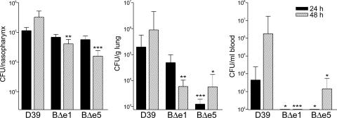
The capacity of Streptococcus pneumoniae to produce capsular polysaccharide (CPS) is essential for virulence. The CPS biosynthesis proteins CpsB, CpsC, and CpsD function to regulate CPS production via tyrosine phosphorylation of CpsD. This mechanism of regulating CPS production is important for enabling S. pneumoniae to cause invasive disease. Here, we identify mutations affecting the attachment of CPS to the cell wall. These mutations were located in cpsC, such that CpsC functioned independently from CpsD tyrosine phosphorylation. These mutants produced WT levels of CPS, but were unable to cause bacteremia in mice after intranasal challenge. This finding suggests that cell-wall attachment of CPS is essential for invasive pneumococcal disease; production of WT levels of CPS alone is not sufficient. We also show that cpsB mutants, which lack the phosphotyrosine-protein phosphatase, produced less CPS than the WT strain, but attached substantially more CPS to their cell wall. Thus, the phosphorylated form of CpsD promotes attachment of CPS to the cell wall.


Conflict of interest statement

Conflict of interest statement: No conflicts declared.

Figures

Fig. 1.
Fig. 1.
Schematic representation of the cpsB mutants. The mutations introduced into various strains are shown schematically as follows: BΔe, cpsB replaced by erm; BΔt, cpsB replaced by tetM; BΔe:DY→F, cpsB replaced by erm and mutation of Y215F, Y218F, Y221F in the [YGX] repeat domain of cpsD; ABΔe, cpsA, and cpsB replaced by erm; AΔe:BNQ, cpsA replaced by erm and mutation of D119N; H201Q in CpsB; BCDΔe1, cpsB, cpsC, and cpsD replaced by erm.
Fig. 2.
Fig. 2.
Comparison of the total amount of CPS produced by various mutants to the amount associated with the cell wall. The amount of total-CPS (A), CW-CPS (B), and THY-CPS (C) was measured, using the uronic acid assay, from (Left to Right) D39, D39-BΔe1, D39-BΔe2, BΔt1, D39-BΔe5, D39-D39-BΔt5, D39-BCDΔe1, D39-AΔ, D39-ABΔe1, D39-AΔe:BNQ2, D39-ABΔe5, D39-AΔe:BNQ6, D39-BΔe:DY→F, D39-DY→F, and D39-DΔ. The results were graphed as percentage compared with D39 total-CPS.
Fig. 3.
Fig. 3.
The number of pneumococci recovered from the nasopharynx, the lung, and the blood after intranasal infection of CD1 mice. The graphs show log10 CFUs (±SE) recovered from 10 CD1 mice for D39, D39-BΔe1 (BΔe1), and BΔe5 (BΔe5) per nasopharynx (total CFUs from nasal wash plus excised nasopharynx, Left), per g of lung tissue (Center), and per ml of blood (Right) after 24 and 48 h. Significant differences when compared with D39, using Student’s unpaired t test (two-tailed), are indicated as follows: ∗∗∗, P < 0.005; ∗∗, P < 0.01; ∗, P < 0.05.
Fig. 4.
Fig. 4.
The effect of cpsB and cpsC mutations on adherence of D39 to A549 cells. The graphs show log10 CFUs (±SE) recovered from each well containing A549 cells infected with D39, D39-BΔe1 (BΔe1), BΔe5 (BΔe5), or D39-DΔ (DΔ). Significant differences when compared with D39, using Student’s unpaired t test (two-tailed), are indicated as follows: ∗∗∗, P < 0.005; ∗∗, P < 0.01; ∗, P < 0.05.

References

    1. Austrian R. Rev. Infect. Dis. 1981;3(Suppl):S1–S17. - PubMed
    1. Henrichsen J. J. Clin. Microbiol. 1995;33:2759–2762. - PMC - PubMed
    1. Magee A. D., Yother J. Infect. Immun. 2001;69:3755–3761. - PMC - PubMed
    1. Selinger D. S., Reed W. P. Infect. Immun. 1979;23:455–458. - PMC - PubMed
    1. Talbot U. M., Paton A. W., Paton J. C. Infect. Immun. 1996;64:3772–3777. - PMC - PubMed

Publication types

MeSH terms

LinkOut - more resources