. 2006 May 17;34(9):2587-97.
doi: 10.1093/nar/gkl280. Print 2006.

Multiple start codons and phosphorylation result in discrete Rad52 protein species

Affiliations

Multiple start codons and phosphorylation result in discrete Rad52 protein species

Adriana Antúnez de Mayolo et al. Nucleic Acids Res. .

Abstract

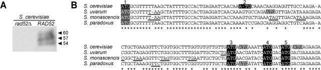
The sequence of the Saccharomyces cerevisiae RAD52 gene contains five potential translation start sites and protein-blot analysis typically detects multiple Rad52 species with different electrophoretic mobilities. Here we define the gene products encoded by RAD52. We show that the multiple Rad52 protein species are due to promiscuous choice of start codons as well as post-translational modification. Specifically, Rad52 is phosphorylated both in a cell cycle-independent and in a cell cycle-dependent manner. Furthermore, phosphorylation is dependent on the presence of the Rad52 C terminus, but not dependent on its interaction with Rad51. We also show that the Rad52 protein can be translated from the last three start sites and expression from any one of them is sufficient for spontaneous recombination and the repair of gamma-ray-induced double-strand breaks.


Figures

Figure 1
Figure 1
Analysis of Rad52 protein. (A) Immunoblot of rad52Δ and RAD52 using anti-Rad52 antibody. Approximate molecular weights (kDa) corresponding to the observed mobility are indicated. (B) Alignment of the RAD52 sequence from four species that belong to the Saccharomyces sensu stricto group: S.cerevisiae, S.paradoxus, S.uvarum and S.monacensis. ATG codons in-frame with the third ATG codon in S.cerevisiae. ATG codons out-of-frame with the third ATG codon in S.cerevisiae. Stop codons in-frame with the previous ATG encountered are underlined. * denotes conserved nucleotides.
Figure 2
Figure 2
Analysis of the third start codon. Note, RAD52-mRNA initiates 10 nt downstream of start codon 2 (15). (A) Schematic diagram of M34A, E36Stop and FS3-4 mutant strains. M34A results from a methionine-to-alanine substitution at amino acid 34. E36Stop results from a stop codon inserted at amino acid 36. FS3-4 mutant results from a frameshift mutation inserted between the third and fourth ATGs. In FS3-4, translation initiated upstream of the fourth start codon results in truncation at amino acid 63. Hence in these mutant strains, Rad52 can only be expressed from the fourth and/or fifth start codons. (B) Schematic diagram of E24Stop M38A M40A mutant having a stop codon at residue 24, and two methionine-to-alanine substitutions at amino acids 38 and 40. In this mutant, translation can only start at the third start codon. M, methionine residues indicating the position of the ATG codons. A, alanine residues substitute the methionine amino acids at the indicated positions. Residues in gray are not productive for translation start. (C) Survival curves of haploid strains after increasing doses of gamma-ray-induced DNA damage. Black diamond, RAD52; open circle, rad52 null; black circle, M34A; black square, FS3-4; inverted open triangle, E36Stop; and open diamond, E24Stop M38A M40A. (D) Gamma-ray survival after overexpression of the RAD52 and M34A genes from 2µ plasmids. Cells carrying 2µ plasmids were grown and plated on SC-His medium. Open square, RAD52 transformed with pRS423 (empty vector); open triangle, M34A transformed with pRS423-M34A; inverted black triangle, M34A transformed with pRS423; and black triangle, M34A transformed with pRS423-RAD52.
Figure 3
Figure 3
Mutational analysis of the fourth and fifth start codons. (A) Schematic diagram of M38A and M40A mutants. M38A or M40A mutant strains result from methionine-to-alanine substitutions at amino acid 38 or 40, respectively. Residues in gray are not productive for translation start. (B) Survival curves of haploid strains after exposure to gamma-irradiation. Black diamond, RAD52; open circle, rad52 null; inverted black triangle, M38A; open triangle, M40A. (C) Schematic diagram of E24Stop M34A M38A and FS4-5 mutants. The E24Stop M34A M38A mutant strain has a stop codon at residue 24 and two methionine-to-alanine substitutions at amino acids 34 and 38. In this triple mutant, translation can only start at the fifth start codon. The FS4-5 mutant results from a frameshift mutation inserted between the fourth and fifth ATGs. In FS4-5, translation initiated upstream of the fifth start codon results in truncation at amino acid 63. M, methionine residues indicating the position of the ATG codons. A, alanine residues substitute the methionine amino acids at the indicated positions. Residues in gray are not productive for translation start. (D) Survival curves of haploid strains after exposure to gamma-irradiation. Black diamond, RAD52; open circle, rad52 null; black circle, FS4-5; and open diamond, E24Stop M34A M38A.
Figure 4
Figure 4
Protein analysis of the rad52 mutants that disrupt the third, fourth and fifth start codons. Rad52 was examined by immunoblot analysis using an anti-Rad52 antibody. Lane 1, rad52 null; lane 2, RAD52; lane 3, E36Stop; lane 4, M34A; lane 5, M38A; lane 6, M40A; lane 7, FS3-4; lane 8, FS4-5; lane 9, E24Stop M38A M40A; and lane 10, E24Stop M34A M38A. Equal amounts of total protein were loaded in each lane on the gel. Approximate molecular weights (kDa) corresponding to the observed mobility are indicated.
Figure 5
Figure 5
Phosphorylation of Rad52. (A) Immunoblot using anti-Rad52 antibody of RAD52 expressed in yeast and E.coli cells. (A–E) Black arrowheads: Rad52 protein bands of different electrophoretic mobilities. (B and C) Gray arrowhead: cell cycle-dependent phosphorylation of Rad52. (B) Cell cycle-independent and cell cycle-dependent phosphorylation of Rad52 protein in E24Stop M38A M40A. Cells were synchronized in G1 by α-factor and released into S phase at time zero. Protein extracts were made from arrested cells (time zero) and at 45 min following α-factor release. (C) Dephosphorylation of Rad52. Wild-type RAD52 cells were synchronized in G1 by α-factor and released into S phase at time zero. Protein extracts were made from arrested cells (time zero) and at 60 min following α-factor release. λ, indicates that protein extracts were treated with λ-phosphatase; −, indicates no phosphatase treatment. (D) Immunoblot analysis of cell cycle synchronized rad52-327delta cells. Protein extracts were made 0, 30 and 60 min after release from G1-arrested cells. Rad52 was examined by immunoblot analysis using anti-Rad52 antibody (I). Samples were subjected to FACS analysis to determine DNA content of cells at each time point (II). (D and E) Empty arrowhead: a protein band non-specifically recognized by the anti-Rad52 antibody also present in the rad52 null. (E) Immunoblot of RAD52 and rad52-409-412delta strains.
Figure 6
Figure 6
Model to explain the multiple Rad52 protein species. (A) RAD52-encoded mRNA allows protein translation from the third and, by leaky scanning, also from the fourth and fifth start codons (3, 4 and 5, respectively). (B) First phosphorylation: cell cycle-independent. Rad52 species from (A) (3, 4 and 5) are modified resulting in slower migrating protein species (3*, 4* and 5*, respectively). (C) Second phosphorylation: cell cycle-dependent. During S phase, some of 3*, 4* and 5* are modified into 3**, 4** and 5**, respectively. Approximate molecular weights (kDa) corresponding to the observed mobility are indicated.

References

    1. Resnick M.A. Genetic control of radiation sensitivity in Saccharomyces cerevisiae. Genetics. 1969;62:519–531. - PMC - PubMed
    1. Game J.C., Mortimer R.K. A genetic study of x-ray sensitive mutants in yeast. Mutat. Res. 1974;24:281–292. - PubMed
    1. Resnick M.A., Martin P. The repair of double-strand breaks in the nuclear DNA of Saccharomyces cerevisiae and its genetic control. Mol. Gen. Genet. 1976;143:119–129. - PubMed
    1. Game J.C., Zamb T.J., Braun R.J., Resnick M., Roth R.M. The role of radiation (rad) genes in meiotic recombination in yeast. Genetics. 1980;94:51–68. - PMC - PubMed
    1. Malone R.E., Esposito R.E. The RAD52 gene is required for homothallic interconversion of mating types and spontaneous mitotic recombination in yeast. Proc. Natl Acad. Sci. USA. 1980;77:503–507. - PMC - PubMed

Publication types

MeSH terms

Substances