. 2006 Jun;188(11):3972-82.
doi: 10.1128/JB.00024-06.

Anatomy of a lactococcal phage tail

Affiliations

Anatomy of a lactococcal phage tail

Stephen Mc Grath et al. J Bacteriol. 2006 Jun.

Abstract

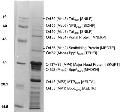
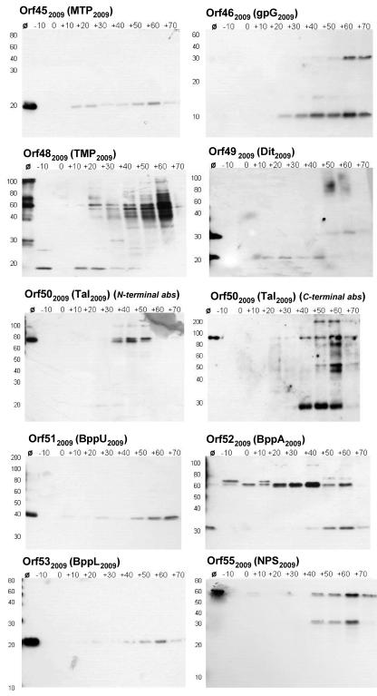
Bacteriophages of the Siphoviridae family utilize a long noncontractile tail to recognize, adsorb to, and inject DNA into their bacterial host. The tail anatomy of the archetypal Siphoviridae lambda has been well studied, in contrast to phages infecting gram-positive bacteria. This report outlines a detailed anatomical description of a typical member of the Siphoviridae infecting a gram-positive bacterium. The tail superstructure of the lactococcal phage Tuc2009 was investigated using N-terminal protein sequencing, Western blotting, and immunogold transmission electron microscopy, allowing a tangible path to be followed from gene sequence through encoded protein to specific architectural structures on the Tuc2009 virion. This phage displays a striking parity with lambda with respect to tail structure, which reenforced a model proposed for Tuc2009 tail architecture. Furthermore, comparisons with lambda and other lactococcal phages allowed the specification of a number of genetic submodules likely to encode specific tail structures.


Figures

FIG. 1.
FIG. 1.
Comparative analysis of the structural tail modules of the three Sfi11-type phages, ul36, Tuc2009, and TP901-1, and the two r1t-type phages, LC3 and r1t. Proteins with predicted or proven functions are indicated by colored arrows. Amino acid identity between proteins is indicated by shaded regions. A schematic representation of genes encoding the structural tail proteins of λ is included for comparison.
FIG. 2.
FIG. 2.
N-terminal sequence analysis of Tuc2009 structural proteins. Concentrated Tuc2009 phage particles were subjected to SDS-PAGE, and the N-terminal sequence of the resulting protein bands was determined. The major structural proteins, MP1, MP2, and MP4, have been reported previously, and their N-terminal sequence is included for consistency. Orf designations and proven or predicted functions are listed opposite the corresponding protein band on the SDS-PAGE gel, and the first five N-terminal amino acids determined are indicated in parentheses.
FIG. 3.
FIG. 3.
TEM analysis of Tuc2009 and model of phage virion showing anatomical features and dimensions. (A) Anatomical features and dimensions of the Tuc2009 virion. (i) Collar and whiskers (NPS2009), (ii) tail shaft (MTP2009), (iii) upper baseplate (BppU2009), (iv) petticoat structure (BppL2009), (v) whiskers (NPS2009), (vi) conical structure (Dit2009), (vii) tail fiber (Tal2009). (B) Detail of proposed protein architecture of tail adsorption apparatus.
FIG. 4.
FIG. 4.
Western blot analysis of Tuc2009 tail proteins. Intracellular protein samples from Tuc2009-infected UC509.9 were probed with polyclonal antibodies specific for individual Tuc2009 tail proteins. Samples were prepared as outlined in Materials and Methods. Polyclonal antibodies used are indicated in the top left corner of each panel, and protein sizes (in kilodaltons) are indicated on the left side of each panel. Ø, CsCl-concentrated Tuc2009 phage particles. −10 to +70, time (in minutes) that a sample was taken, relative to the addition of phage at time zero.
FIG. 5.
FIG. 5.
Immunogold TEM analysis of Tuc2009. Specific proteins were located on the Tuc2009 and TP901-1 (48 mutant) virions as described in Materials and Methods. Panels A to F, Tuc2009. Panel G, TP901-1. (A) Anti-45abs (MTP2009), (B) anti-49abs (Dit2009), (C) anti-50Cabs (Tal2009), (D) anti-51abs (BppU2009), (E) anti-53abs (BppL2009), (F) anti-55abs (NPS), (G) anti-48abs (TMP2009).

References

    1. Ackermann, H. W. 1996. Frequency of morphological phage descriptions in 1995. Arch. Virol. 141:209-218. - PubMed
    1. Arendt, E. K., C. Daly, G. F. Fitzgerald, and M. van de Guchte. 1994. Molecular characterization of lactococcal bacteriophage Tuc2009 and identification and analysis of genes encoding lysin, a putative holin, and two structural proteins. Appl. Environ. Microbiol. 60:1875-1883. - PMC - PubMed
    1. Blatny, J. M., L. Godager, M. Lunde, and I. F. Nes. 2004. Complete genome sequence of the Lactococcus lactis temperate phage phiLC3: comparative analysis of phiLC3 and its relatives in lactococci and streptococci. Virology 318:231-244. - PubMed
    1. Braun, V., Jr., S. Hertwig, H. Neve, A. Geis, and M. Teuber. 1989. Taxonomic differentiation of bacteriophages of Lactococcus lactis by electron microscopy, DNA-DNA hybridization and protein profiles. J. Gen. Microbiol. 181:7291-7297.
    1. Brøndsted, L., S. Østergaard, M. Pedersen, K. Hammer, and F. K. Vogensen. 2001. Analysis of the complete DNA sequence of the temperate bacteriophage TP901-1: evolution, structure, and genome organization of lactococcal bacteriophages. Virology 283:93-109. - PubMed

Publication types