. 2006 May 17;26(20):5500-10.
doi: 10.1523/JNEUROSCI.3955-05.2006.

Urocortin 2-deficient mice exhibit gender-specific alterations in circadian hypothalamus-pituitary-adrenal axis and depressive-like behavior

Affiliations

Urocortin 2-deficient mice exhibit gender-specific alterations in circadian hypothalamus-pituitary-adrenal axis and depressive-like behavior

Alon Chen et al. J Neurosci. .

Abstract

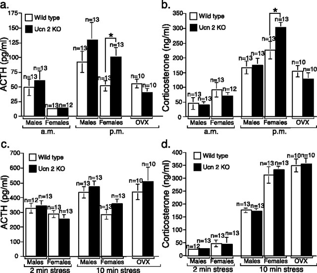
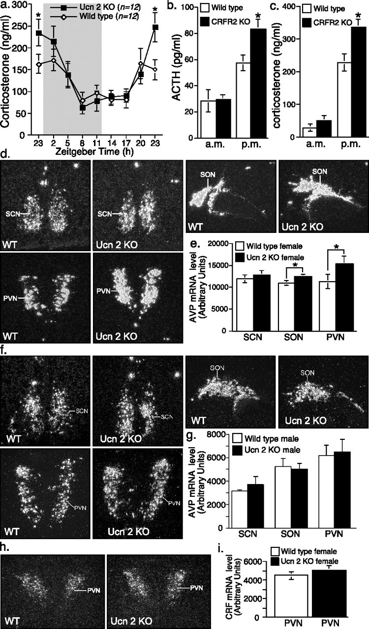
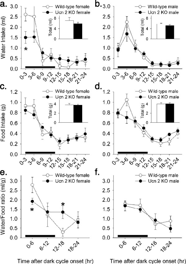
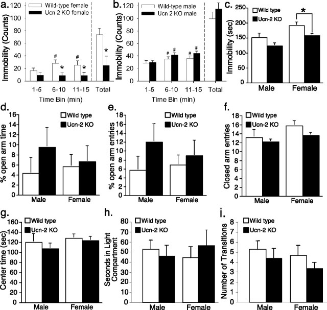
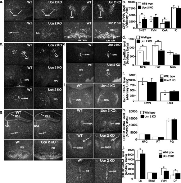
Gender differences in hypothalamus-pituitary-adrenal (HPA) axis activation and the prevalence of mood disorders are well documented. Urocortin 2, a recently identified member of the corticotropin-releasing factor family, is expressed in discrete neuroendocrine and stress-related nuclei of the rodent CNS. To determine the physiological role of urocortin 2, mice null for urocortin 2 were generated and HPA axis activity, ingestive, and stress-related behaviors and alterations in expression levels of CRF-related ligands and receptors were examined. Here we report that female, but not male, mice lacking urocortin 2 exhibit a significant increase in the basal daily rhythms of ACTH and corticosterone and a significant decrease in fluid intake and depressive-like behavior. The differential phenotype of urocortin 2 deficiency in female and male mice may imply a role for urocortin 2 in these gender differences.


Figures

Figure 1.
Figure 1.
For clarity, cosinor measures (e.g., MESOR, amplitude, and acrophase) of circadian regulation of drug and food intake are illustrated in a sample figure that depicts a cosinor function with MESOR of 15 h, amplitude of 10 h, peak of 25 h, nadir of 5 h, and acrophase of 13 h from a reference point (e.g., dark onset).
Figure 2.
Figure 2.
Generation of Ucn-2-deficient mice. a, Structure of the Ucn-2 locus (top), targeting vector used to transfect ES cells (middle), and the mutated locus (bottom). b, Southern blot and PCR analysis of tail DNA isolated from the progeny of a heterozygote cross (wild-type, +/+; heterozygous, +/−; and null mutant, −/−). c, RT-PCR assay demonstrated the absence of Ucn-2 mRNA in the brain, skeletal muscle, and skin of the Ucn-2 mutant mice. d, Immunohistochemistry demonstrated the absence of Ucn-2 protein in the locus ceruleus (LC) of Ucn-2 mutant mice. 4V, Fourth ventricle.
Figure 3.
Figure 3.
Higher nocturnal basal (prestress) ACTH and corticosterone levels in female Ucn-2 null mutant mice. a, b, Basal (prestress) ACTH (a) and corticosterone (b) plasma levels at 7:00 A.M. (a.m.) and 5:00 P.M. (p.m.) in male and female Ucn-2 mutant mice and WT littermates. c, d, Plasma concentrations of ACTH (c) and corticosterone (d) in Ucn-2 null mice and WT littermates after 2 or 10 min of restraint stress. Note that Ucn-2 mutant mice show normal hormonal responses to acute restraint stress. Data are displayed as the mean ± SEM. *p < 0.05 vs WT mice.
Figure 4.
Figure 4.
Elevated corticosterone levels in female Ucn-2 null mice during the light/dark cycle transition interphase and increase in hypothalamic AVP expression. a, A 24 h corticosterone profile for female Ucn-2 null and WT mice showed elevated levels in mutant females during the interphase light/dark cycle transition. Gray shading indicates the time of lights off. b, c, Basal (prestress) ACTH (b) and corticosterone (c) plasma levels at 7:00 A.M. (a.m.) and 5:00 P.M. (p.m.) in female CRFR2 mutant mice and WT littermates. Elevated ACTH and corticosterone levels were observed during the evening sampling. d–g, Representative dark-field photomicrographs (d, f) and semiquantitative analysis (e, g) showing in situ hybridization for hypothalamic AVP in WT and Ucn-2 null female (d, e) or male (f, g) mice. The AVP mRNA expression in the hypothalamic SON and PVN are significantly increased in mutant females, but not male, compared with WT littermates mice. h, i, Representative dark-field photomicrographs (h) and semiquantitative analysis (i) showing in situ hybridization for CRF in the PVN of WT and Ucn-2 null female mice. Note that Ucn-2 mutant mice show no significant differences in CRF mRNA expression in the PVN compared with WT littermates. SCN, Suprachiasmatic nucleus; *p < 0.05 vs WT mice.
Figure 5.
Figure 5.
Blunted circadian amplitude of spontaneous water intake in female Ucn-2 mutant mice. a, b, A 24 h profile of water intake showed decreased peak water intake at the onset of the dark cycle and a trend for increased water intake at the diurnal nadir in female, but not male, Ucn-2 null mice. c, d, In contrast, the circadian profile of eating was not reliably altered in Ucn-2 null mutant mice of either sex. e, f, Female, but not male, Ucn-2 null mutant mice showed a flattening of the circadian rhythm of the water/food ratio, in which rodents typically drink in greater quantities relative to food during the nocturnal compared with diurnal cycles. Split-plot ANOVA revealed significant genotype × time interactions for the time course of drinking (F(7,70) = 2.17; p < 0.05) and water/food ratios (p < 0.05) in female, but not male, mice. Table 1 further quantifies differences in the circadian profile of ingestion using cosinor analysis. Data are displayed as the mean ± SEM, and the black bar represents the dark cycle. *p < 0.05 vs WT condition (Student’s t test).
Figure 6.
Figure 6.
Altered effect of d-fenfluramine on food intake in Ucn-2 null mice. a, c, Pretreatment with d-fenfluramine (2 mg/kg, i.p.) produced a characteristic acute hypophagic response in WT mice that was followed by hyperphagia, perhaps compensatory, relative to vehicle (10 ml/kg 0.9% saline) conditions. b, d, Ucn-2 null mutant mice did not exhibit the delayed post-fenfluramine hyperphagia, and female null mutant mice exhibited a smaller hypophagic response than WT mice. Effects were reflected in significant genotype × drug and genotype × drug × time interactions. Data are expressed as the mean ± SEM. *p < 0.05 versus vehicle (post hoc within-subject Student’s t test).
Figure 7.
Figure 7.
Increased antidepressant-like, but not anxiolytic-like, behavior in female Ucn-2 null mutant mice. a, b, Female Ucn-2 null mice did not become increasingly immobile during a modified forced-swim test, unlike female WT mice and males of both genotypes. Behavioral analysis indicated that this resulted mainly from persistent swimming, a behavior linked to serotonergic acting antidepressants (see Results). c, Female Ucn-2 null mutant mice also were significantly less immobile in the tail-suspension test. Ucn-2 null mutant mice did not differ in measures of anxiety-like behavior in the elevated plus maze (d, e) or light/dark box (h) tests, nor did they differ in measures related to locomotor/exploratory activity during the elevated plus maze (f, g) or light/dark box (i) tests. Data are displayed as the mean ± SEM. *p < 0.05 versus WT mice (main effect of ANOVA for forced swim, Student’s t test for tail suspension); #p < 0.05 versus 1–5 min time bin (within-subject Dunnett’s test).
Figure 8.
Figure 8.
Locomotor activity and rearing behavior and contextual and cued conditioning in Ucn-2 null and WT mice. A–d, Twenty-eight hour horizontal (locomotor; a, b) activity or vertical (rearing; c, d) behavior for Ucn-2 null and WT female (a, c) or male (b, d) mice. Data are displayed as the mean ± SEM. The black bar represents the dark cycle. e–l, All four trials are shown (habituation, e, f; conditioning, g, h; context test, i, j, and conditioned stimulus, k, l) for male (e, g, i, k) and female (f, h, j, l) mice. The arrows on the conditioning trial graphs represent the onset of the 2 s shock. Data are expressed as the mean ± SEM of the time (seconds) spent freezing within the time interval indicated on the horizontal axis.
Figure 9.
Figure 9.
Increased CRF and CRFR2 and decreased Ucn-3 mRNA expression in the brain of Ucn-2 mutant mice. a, b, Representative dark-field photomicrographs (a) and semiquantitative analysis (b) showing in situ hybridization for CRF in WT and Ucn-2 null mice brain. The CRF mRNA expression in the BNST and CeA are significantly increased in mutant mice. c, d, Representative dark-field photomicrographs (c) and semiquantitative analysis (d) showing in situ hybridization for Ucn-3 in WT and Ucn-2 null mice brain. The Ucn-3 mRNA expression in the MPO and PeF are significantly decreased in mutant mice. e, f, Representative dark-field photomicrographs (e) and semiquantitative analysis (f) showing in situ hybridization for Ucn-1 in WT and Ucn-2 null mice brain. Note that Ucn-2 mutant mice show no significant differences in Ucn-1 mRNA expression in the brain compared with WT littermates. g, h, Representative dark-field photomicrographs (g) and semiquantitative analysis (h) showing in situ hybridization for CRFR1 in WT and Ucn-2 null mice brain. Note that Ucn-2 mutant mice show no significant differences in CRFR1 mRNA expression in the brain compared with WT littermates. i, j, Representative dark-field photomicrographs (i) and semiquantitative analysis (j) showing in situ hybridization for CRFR2 in WT and Ucn-2 null mice brain. The CRFR2 mRNA expression in the LS, ventral medial hypothalamus, and DR are significantly increased in mutant mice. IO, Inferior olive; MeA, medial amygdala; EWN, Edinger Westphal nucleus; LSO, lateral superior olive; VMH, ventral medial hypothalamus; HPC, hippocampus, RT, reticular nucleus; PG, pontine gray.

Similar articles

Cited by

References

    1. Amat J, Tamblyn JP, Paul ED, Bland ST, Amat P, Foster AC, Watkins LR, Maier SF (2004). Microinjection of urocortin 2 into the dorsal raphe nucleus activates serotonergic neurons and increases extracellular serotonin in the basolateral amygdala. Neuroscience 129:509–519. - PubMed
    1. Arborelius L, Owens MJ, Plotsky PM, Nemeroff CB (1999). The role of corticotropin-releasing factor in depression and anxiety disorders. J Endocrinol 160:1–12. - PubMed
    1. Bale TL, Vale WW (2003). Increased depression-like behaviors in corticotropin-releasing factor receptor-2-deficient mice: sexually dichotomous responses. J Neurosci 23:5295–5301. - PMC - PubMed
    1. Bale TL, Vale WW (2004). CRF and CRF receptors: role in stress responsivity and other behaviors. Annu Rev Pharmacol Toxicol 44:525–557. - PubMed
    1. Bale TL, Contarino A, Smith GW, Chan R, Gold LH, Sawchenko PE, Koob GF, Vale WW, Lee KF (2000). Mice deficient for corticotropin-releasing hormone receptor-2 display anxiety-like behaviour and are hypersensitive to stress. Nat Genet 24:410–414. - PubMed

Publication types

MeSH terms

LinkOut - more resources