. 2006 May 17;26(20):5554-64.
doi: 10.1523/JNEUROSCI.5156-05.2006.

Early life blockade of 5-hydroxytryptamine 1A receptors normalizes sleep and depression-like behavior in adult knock-out mice lacking the serotonin transporter

Affiliations

Early life blockade of 5-hydroxytryptamine 1A receptors normalizes sleep and depression-like behavior in adult knock-out mice lacking the serotonin transporter

Chloé Alexandre et al. J Neurosci. .

Abstract

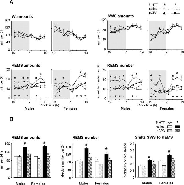
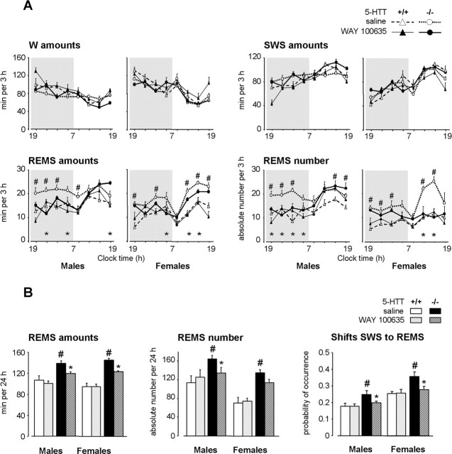
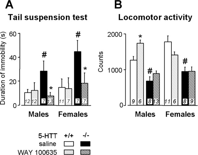
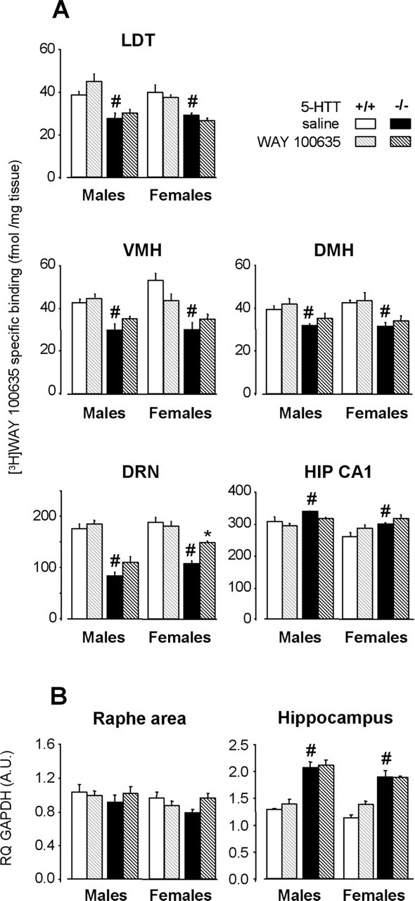
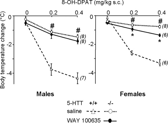
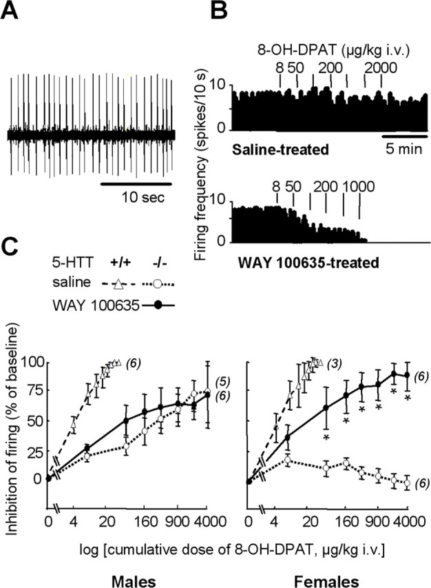
In serotonin transporter knock-out (5-HTT-/-) mice, extracellular serotonin (5-HT) levels are markedly elevated in the brain, and rapid eye movement sleep (REMS) is enhanced compared with wild-type mice. We hypothesized that such sleep impairment at adulthood results from excessive serotonergic tone during early life. Thus, we assessed whether neonatal treatment with drugs capable of limiting the impact of 5-HT on the brain could normalize sleep patterns in 5-HTT-/- mutants. We found that treatments initiated at postnatal day 5 and continued for 2 weeks with the 5-HT synthesis inhibitor para-chlorophenylalanine, or for 4 weeks with the 5-HT(1A) receptor (5-HT(1A)R) antagonist N-[2-[4-(2-methoxyphenyl)-1-piperazinyl]ethyl]-N-(2-pyridinyl) cyclohexane carboxamide (WAY 100635), induced total or partial recovery of REMS, respectively, in 5-HTT-/- mutants. Early life treatment with WAY 100635 also reversed the depression-like behavior otherwise observed in these mutants. Possible adaptive changes in 5-HT(1A)R after neonatal treatment with WAY 100635 were investigated by measuring 5-HT(1A) binding sites and 5-HT(1A) mRNA in various REMS- and/or depression-related brain areas, as well as 5-HT(1A)R-mediated hypothermia and inhibition of neuronal firing in the dorsal raphe nucleus. None of these characteristics were modified in parallel with REMS recovery, suggesting that 5-HT(1A)Rs involved in wild-type phenotype rescue in 5-HTT-/- mutants are located in other brain areas or in 5-HT(1A)R-unrelated circuits where they could be transiently expressed during development. The reversal of sleep alterations and depression-like behavior after early life blockade of 5-HT(1A)R in 5-HTT-/- mutants might open new perspectives regarding preventive care of sleep and mood disorders resulting from serotonin transporter impairments during development.


Figures

Figure 1.
Figure 1.
Effects of a 2 week neonatal treatment with pCPA on sleep–wakefulness patterns in 5-HTT−/− and 5-HTT+/+ mice. A, Sleep–wakefulness states across the light/dark cycle in male (left) and female (right) 5-HTT−/− (bold lines) and 5-HTT+/+ (thin lines) mice treated neonatally with saline (open symbols, dotted lines) or pCPA (filled symbols, solid lines). The data are expressed as minutes per 3 h for the amounts of vigilance states and as absolute value for the number of REMS episodes. B, REMS amounts, number of episodes, and number of shifts from SWS to REMS (expressed as probability of occurrence normalized to total number of shifts from SWS) are calculated for 24 h, in male (left) and female (right) 5-HTT−/− (black bars) and 5-HTT+/+ (grey bars) mice treated with saline (solid bars) or pCPA (hatched bars). All data are expressed as means ± SEM of three to nine animals per group (see Table 1 for the number of animals in each group). *p < 0.05, significant difference for treatment (Fisher’s test). #p < 0.05, significant difference for genotype in mice treated with saline (Fisher’s test).
Figure 2.
Figure 2.
Effects of a 4 week neonatal treatment with WAY 100635 on sleep–wakefulness patterns in 5-HTT−/− and 5-HTT+/+ mice. A, Sleep–wakefulness states across the light/dark cycle in male (left) and female (right) 5-HTT−/− (bold lines) and 5-HTT+/+ (thin lines) mice treated neonatally with saline (open symbols, dotted lines) or WAY 100635 (filled symbols, solid lines). The data are expressed as minutes per 3 h for the amounts of vigilance states and as absolute value for the number of REMS episodes. B, REMS amounts, number of episodes, and number of shifts from SWS to REMS (expressed as probability of occurrence normalized to total number of shifts from SWS) are calculated for 24 h, in male (left) and female (right) 5-HTT−/− (black bars) and 5-HTT+/+ (grey bars) mice treated with saline (solid bars) or WAY 100635 (hatched bars). All data are expressed as means ± SEM of four to seven animals per group (see Table 1 for the number of animals in each group). *p < 0.05, significant difference for treatment (Fisher’s test). #p < 0.05, significant difference for genotype in mice treated with saline (Fisher’s test).
Figure 3.
Figure 3.
Effects of a 4 week neonatal treatment with WAY 100635 on behavior in the tail suspension test (A) and on locomotor activity in a novel environment (B), in male (left) and female (right) 5-HTT−/− (black bars) and 5 HTT+/+ (grey bars) mice treated with saline (solid bars) or WAY 100635 (hatched bars). Data are expressed as means ± SEM for the number of animals indicated in bars. *p < 0.05, significant difference for treatment (Fisher’s test). #p < 0.05, significant difference for genotype in mice treated with saline (Fisher’s test).
Figure 4.
Figure 4.
5-HT1AR labeling and mRNA levels after a 4 week neonatal treatment with WAY 100635 (hatched bars) or saline (solid bars) in male (left) and female (right) 5-HTT−/− (black bars) and 5-HTT+/+ (grey bars) mice. A, Quantitative autoradiography of 5-HT1AR labeling by [3H]WAY 100635 in various brain areas. Data (mean ± SEM of 3–4 animals per group) are expressed as femtomoles per milligram of tissue. B, 5-HT1AR mRNA levels in the anterior raphe area and the hippocampus. Results [relative quantity (RQ); mean ± SEM of 4–5 animals per group] are expressed as arbitrary units (A.U.) after normalization to an endogenous reference gene (GAPDH). Normalization with HPRT endogenous gene (see Materials and Methods) yielded similar results. LDT, Laterodorsal tegmental nucleus; VMH, ventromedial nucleus of hypothalamus; DMH, dorsomedial nucleus of hypothalamus; HIP CA1, field of hippocampus. *p < 0.05, significant difference for treatment (Fisher’s test). #p < 0.05, significant difference for genotype in mice treated with saline (Fisher’s test).
Figure 5.
Figure 5.
Dose-dependent 8-OH-DPAT-induced hypothermia in male (left) and female (right) 5-HTT−/− mutant mice after early life treatment with saline (open symbols, dotted lines) or WAY 100635 (filled symbols, solid lines). Core body temperature changes (mean ± SEM for the number of animals indicated in parentheses), expressed as degrees Celsius, are the difference between baseline and the lowest temperature recorded within 60 min after subcutaneous injection of saline (0 on abscissa) or 8-OH-DPAT (0.2 and 0.4 mg/kg). *p < 0.05, significant difference for treatment (Fisher’s test). For reference data, 8-OH-DPAT-induced hypothermia was also measured in groups of paired wild-type mice that had received neonatal treatment with saline (open triangles, dashed lines). #p < 0.05, significant difference for genotype in mice treated with saline (Fisher’s test).
Figure 6.
Figure 6.
Functional status of 5-HT1A autoreceptors in the DRN. A, Oscilloscope trace showing spontaneous firing activity of a 5-HT neuron recorded in the DRN of a wild-type female mouse. B, Examples of integrated firing rate (in spikes per 10 s) histograms from female 5-HTT−/− mutants treated neonatally with saline (top) or WAY 100635 (bottom). Vertical bars above histograms represent injections of 8-OH-DPAT at the dose indicated (in μg/kg, i.v.). C, Dose-dependent inhibition by 8-OH-DPAT of DRN neuron firing in male (left) and female (right) 5-HTT−/− mutant mice after neonatal treatment with saline (open symbols, dotted lines) or WAY 100635 (filled symbols, solid lines). Inhibition of neuron firing (mean ± SEM of the number of neurons indicated in parentheses) is expressed as a percentage of baseline. Firing frequency was measured during the second minute after intravenous injection of cumulative doses of 8-OH-DPAT (on abscissa). The estrous cycle was not monitored because it does not influence this neuronal response (Bouali et al., 2003). *p < 0.05, significant difference for treatment (Fisher’s test). For reference data, 8-OH-DPAT-induced inhibition of neuronal firing was also assessed in groups of paired wild-type mice that had received neonatal treatment with saline (open triangles, dashed lines).

References

    1. Adrien J (2002). Neurobiological bases for the relation between sleep and depression. Sleep Med Rev 6:351–352. - PubMed
    1. Alexandre C, Popa D, Bouali S, Léna C, Hamon M, Adrien J (2003). Inhibition of serotonin synthesis during development restores normal REM sleep level in 5-HT transporter knock-out mice. Sleep 26:[Abstr Suppl], 0071A.
    1. Amici R, Sanford LD, Kearney K, McInerney B, Ross RJ, Horner RL, Morrison AR (2004). A serotonergic (5-HT2) receptor mechanism in the laterodorsal tegmental nucleus participates in regulating the pattern of rapid-eye-movement sleep occurrence in the rat. Brain Res 996:9–18. - PubMed
    1. Ansorge MS, Zhou M, Lira A, Hen R, Gingrich JA (2004). Early-life blockade of the 5-HT transporter alters emotional behavior in adult mice. Science 306:879–881. - PubMed
    1. Beique JC, Campbell B, Perring P, Hamblin MW, Walker P, Mladenovic L, Andrade R (2004). Serotonergic regulation of membrane potential in developing rat prefrontal cortex: coordinated expression of 5-hydroxytryptamine (5-HT)1A, 5-HT2A, and 5-HT7 receptors. J Neurosci 24:4807–4817. - PMC - PubMed

Publication types

MeSH terms

Substances