. 2006 May 30;103(22):8517-22.
doi: 10.1073/pnas.0602657103. Epub 2006 May 18.

Structural and functional recovery from early monocular deprivation in adult rats

Affiliations

Structural and functional recovery from early monocular deprivation in adult rats

Tommaso Pizzorusso et al. Proc Natl Acad Sci U S A. .

Abstract

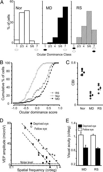
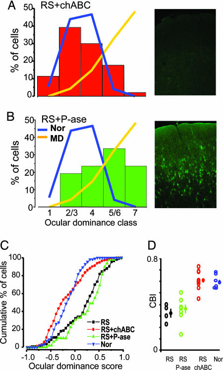
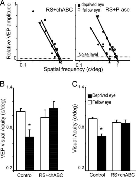
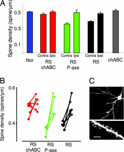
Visual deficits caused by abnormal visual experience during development are hard to recover in adult animals. Removal of chondroitin sulfate proteoglycans from the mature extracellular matrix with chondroitinase ABC promotes plasticity in the adult visual cortex. We tested whether chondroitinase ABC treatment of adult rats facilitates anatomical, functional, and behavioral recovery from the effects of a period of monocular deprivation initiated during the critical period for monocular deprivation. We found that chondroitinase ABC treatment coupled with reverse lid-suturing causes a complete recovery of ocular dominance, visual acuity, and dendritic spine density in adult rats. Thus, manipulations of the extracellular matrix can be used to promote functional recovery in the adult cortex.


Conflict of interest statement

Conflict of interest statement: No conflicts declared.

Figures

Fig. 1.
Fig. 1.
Persistence of MD effects in adult RS rats. (A) The ocular dominance of visual cortical neurons remains shifted toward the previously nondeprived eye in adult RS rats. Ocular dominance distributions of nondeprived [normal (Nor); n = 6; 149 cells], long-term MD (MD; n = 5; 95 cells), and RS (RS; n = 4; 80 cells) rats significantly differ from each other (χ2 test, P < 0.05). (B) Ocular dominance score distributions of Nor, MD, and RS rats (Kolmogorov–Smirnov test, P < 0.05). (C) CBIs (open circles, values of single animals; filled circles, mean ± SE) of Nor, MD, and RS rats (one-way ANOVA, P < 0.001; all groups differ form each other; post hoc Tukey test, P < 0.05). (D) Representative example of visual acuity estimates of both eyes in an RS animal. VEP transient amplitude is plotted against the spatial frequency log. Visual acuity is taken as the spatial frequency that coincides with the extrapolation to zero amplitude of the linear regression through the last four to five data points above noise level. Dashed lines indicate noise level. (E) The visual acuity of the formerly deprived eye is significantly lower than that of the other eye in both MD and RS adult rats (paired t test, P < 0.05). Visual acuity of the other eye was within the normal acuity values estimated with VEPs or behaviorally in the rat.
Fig. 2.
Fig. 2.
chABC treatment allows complete recovery of ocular dominance in adult RS rats. (A Left) Ocular dominance distribution of RS chABC-treated (RS+chABC; n = 9; 204 cells) rats is statistically undistinguishable from that of adult normal rats (χ2 test, P > 0.05). (A Right) Perineuronal and diffuse WFA staining is completely abolished in visual cortex of recorded chABC-treated animals. (B Left) Ocular dominance distribution of adult RS P-ase-treated (RS+P-ase; n = 7; 140 cells) rats remains significantly shifted toward the previously deprived eye. For comparison, the distribution of normal adults (Nor, blue line) and long-term MD rats (MD, orange line) reported in Fig. 1 is outlined. Data for the Nor and MD groups are the same as those in Fig. 1. (B Right) Normal appearance of CSPG-containing PNNs stained with WFA in a recorded rat treated with P-ase (field centered on layer 4). (C) The ocular dominance score distribution of RS+chABC animals is significantly shifted toward normal values with respect to both the RS and RS+P-ase groups. The RS and RS+P-ase groups do not differ between themselves (Kolmogorov–Smirnov statistics, level of significance of 0.05). (D) CBIs (open circles, values of single animals; filled circles, mean ± SE) of RS, RS+P-ase, RS+chABC, and Nor rats. Statistical analysis shows that CBIs of RS+chABC rats are not different from those of normal rats but differ from CBIs of RS and RS+P-ase rats (one-way ANOVA, level of significance P = 0.001; all groups differ from each other; post hoc Tukey test, level of significance P = 0.05).
Fig. 3.
Fig. 3.
chABC causes a complete recovery of visual acuity of the previously deprived eye. (A) Representative examples of visual acuity assessment of the previously deprived and nondeprived eyes in two rats treated with P-ase (Right) and chABC (Left) after 1 week of RS. VEP amplitude is normalized to peak amplitude. (B) Visual acuity of the two eyes in both experimental groups. Statistical analysis confirms that the treatment with chABC promoted a complete recovery of visual acuity of the formerly deprived eye (paired t test, level of significance of 0.05). (C) Behavioral visual acuity of a normal eye (open bar) and long-term-deprived eye (filled bar) of control animals [n = 4 (two P-ase rats and two untreated rats)] and chABC-treated animals (n = 4). Paired t test shows that visual acuity of the long-term-deprived eye is significantly reduced with respect to the normal eye in controls but not in chABC-treated animals. Asterisks in B and C indicate statistically different groups.
Fig. 4.
Fig. 4.
chABC combined with RS causes a significant recovery of dendritic spine density. (A) Mean spine density in normal rats (Nor), RS rats, RS P-ase-treated rats (RS P-ase), RS chABC-treated rats (RS chABC), and nondeprived rats treated with chABC (chABC). The data of RS, RS P-ase, and RS chABC are reported separately for the cortex ipsilateral (Ipsi) to the long-term-deprived eye that is untreated and the cortex contralateral (Contra) to the long-term-deprived eye that is treated with chABC or P-ase. In both RS P-ase and RS rats, spine density in the cortex contralateral to the deprived eye is smaller than in normal rats. In contrast, spine density in RS chABC-treated rats did not differ from normal rats (one-way ANOVA, P < 0.001; post hoc Tukey test, Nor vs. RS and RS P-ase, P < 0.001; Nor vs. RS chABC, P = 0.4). Spine density in the cortex ipsilateral to the deprived eye was not different among groups (Nor, RS, RS P-ase, RS chABC; one-way ANOVA, P = 0.82). The chABC treatment per se does not affect spine density (chABC vs. Nor, P = 0.42, t test). (B) Within-animal comparison of spine density in RS, RS P-ase, and RS chABC. Data from single animals are reported. Filled circles represent data for the cortex contralateral to the long-term-deprived eye; open circles represent values for the cortex ipsilateral to the long-term-deprived eye. Data from the same animal are connected by a segment. In RS and RS P-ase, density is significantly smaller in the contralateral cortex (RS P-ase, P = 0.037; RS, P = 0.003; paired t test). No difference is present between the two cortices of RS chABC rats (P = 0.29, paired t test). (C) Example of a layer II–III pyramid cell of the binocular visual cortex and of a dendritic branch. (Scale bar in C: Upper, 40 μm; Lower, 5 μm.)

References

    1. Mitchell D. E., MacKinnon S. Clin. Exp. Optom. 2002;85:5–18. - PubMed
    1. Hubel D. H., Wiesel T. N. J. Physiol. (London) 1970;206:419–436. - PMC - PubMed
    1. Fagiolini M., Pizzorusso T., Berardi N., Domenici L., Maffei L. Vision Res. 1994;34:709–720. - PubMed
    1. Mataga N., Mizuguchi Y., Hensch T. K. Neuron. 2004;44:1031–1041. - PubMed
    1. Oray S., Majewska A., Sur M. Neuron. 2004;44:1021–1030. - PubMed

Publication types

Substances