. 2006 May 30;103(22):8493-8.
doi: 10.1073/pnas.0509821103. Epub 2006 May 18.

A unique strategy for mRNA cap methylation used by vesicular stomatitis virus

Affiliations

A unique strategy for mRNA cap methylation used by vesicular stomatitis virus

Jianrong Li et al. Proc Natl Acad Sci U S A. .

Abstract

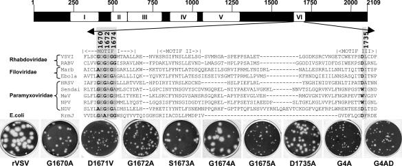
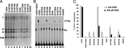
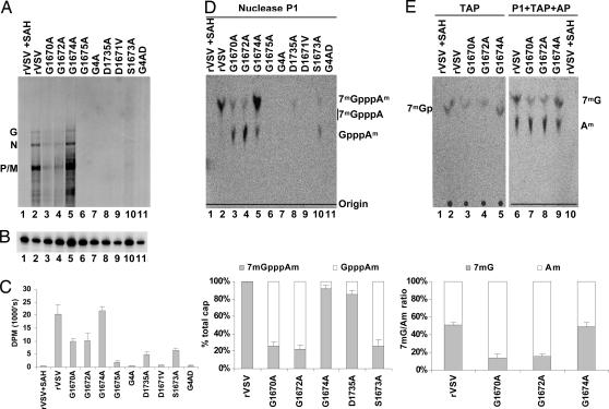
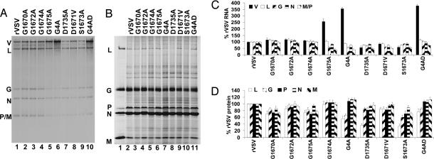
Nonsegmented negative-sense (nsNS) RNA viruses typically replicate within the host cell cytoplasm and do not have access to the host mRNA capping machinery. These viruses have evolved a unique mechanism for mRNA cap formation in that the guanylyltransferase transfers GDP rather than GMP onto the 5' end of the RNA. Working with vesicular stomatitis virus (VSV), a prototype nsNS RNA virus, we now provide genetic and biochemical evidence that its mRNA cap methylase activities are also unique. Using recombinant VSV, we determined the function in mRNA cap methylation of a predicted binding site in the polymerase for the methyl donor, S-adenosyl-l-methionine. We found that amino acid substitutions to this site disrupted methylation at the guanine-N-7 (G-N-7) position or at both the G-N-7 and ribose-2'-O (2'-O) positions of the mRNA cap. These studies provide genetic evidence that the two methylase activities share an S-adenosyl-l-methionine-binding site and show that, in contrast to other cap methylation reactions, methylation of the G-N-7 position is not required for 2'-O methylation. These findings suggest that VSV evolved an unusual strategy of mRNA cap methylation that may be shared by other nsNS RNA viruses.


Conflict of interest statement

Conflict of interest statement: No conflicts declared.

Figures

Fig. 1.
Fig. 1.
AdoMet-binding site alterations. (Upper) Amino acid sequence alignments of a predicted AdoMet-binding region of domain VI of nsNS RNA virus L proteins and known RNA methylases. The conserved motifs of nsNS RNA virus polymerases (I–VI) are shown (28). The AdoMet-binding residues modified in this study are shaded. VSVI, VSV Indiana; RABV, rabies virus; Marb, Marburg; HRSV, human respiratory syncytial virus; MeV, measles virus; NPV, Nipah virus; NDV, Newcastle disease virus; RrmJ, Escherichia coli 2′-O MTase. (Lower) Plaque morphology of recombinant viruses on Vero cells. Plaques of rVSV and G1674A were developed after 24 h; those of G1670A, G1672A, G1675A, D1735A, D1671V, and S1673A were developed after 48 h; those of G4A and G4AD were developed after 96 h.
Fig. 2.
Fig. 2.
Effect of L gene mutations on G-N-7 methylation. (A) Transcription reactions were performed in the presence of [α-32P]GTP, RNA was analyzed by electrophoresis on acid-agarose gels, and products were detected by using a phosphoimager. The virus and the migration of the RNA are shown. (B) RNA was synthesized in the presence of 200 μM AdoMet or SAH and 15 μCi of [α-32P]GTP and digested with 2 units of TAP, and the products were analyzed by TLC on PEI cellulose F sheets. Plates were dried, and the spots were visualized with a phosphoimager. The migration of the markers 7mGp and Gp are shown. (C) Quantitative analysis of three independent experiments. For each virus, the released 7mGp (mean ± SD) was expressed as a percentage of the total released cap structure.
Fig. 3.
Fig. 3.
Effect of L gene mutations on 2′-O and G-N-7 methylation. (A) RNA was synthesized in the presence of [3H]AdoMet and analyzed by electrophoresis on acid-agarose gels. The virus and the identity of the mRNAs are shown. (B) RNA from A was examined by primer extension assay by using a primer designed to anneal to the N mRNA. (C) [3H]AdoMet incorporation monitored by scintillation counting. Three independent experiments were used to generate the graph shown. (D and E) (Upper) RNA was digested with P1, TAP, and AP, and the products were analyzed by TLC on PEI cellulose F sheets. Plates were dried, and the spots were visualized with a phosphoimager. The identity of the virus and the migration of the markers 7mGpppA, GpppA, 7mG, and 2′-OmA are shown. (Lower) Quantitative analysis of three independent experiments is shown. For each virus, the fraction of the mRNA cap that was 7mGpppAm and GpppAm or 7mG and 2′-OmA is shown (mean ± SD).
Fig. 4.
Fig. 4.
Effect of L gene mutations on viral gene expression in BHK-21 cells. (A) Cells were infected at an moi of 3, and RNAs were labeled with [3H]uridine, resolved by electrophoresis on acid-agarose gels, and visualized by fluorography. RNA extracted from an equivalent number of cells was loaded in each lane. The virus and the identity of the RNAs are shown. V, replication products; L, G, N, and P/M, mRNA. (B) Proteins were labeled by incorporation of [35S]Express, and cytoplasmic extracts were analyzed by SDS/PAGE and detected by using a phosphoimager. Extract from equivalent numbers of cells was loaded in each lane. The virus and the identity of the proteins are shown. (C and D) Quantitative analysis of RNA (C) and protein abundance (D). The mean ± SD was expressed as a percentage of that observed for rVSV from three independent experiments.

References

    1. Muthukrishnan S., Morgan M., Banerjee A. K., Shatkin A. J. Biochemistry. 1976;15:5761–5768. - PubMed
    1. Furuichi Y., LaFiandra A., Shatkin A. J. Nature. 1977;266:235–239. - PubMed
    1. Furuichi Y., Shatkin A. J. Adv. Virus Res. 2000;55:135–184. - PMC - PubMed
    1. Shuman S. Prog. Nucleic Acid Res. Mol. Biol. 2001;66:1–40. - PubMed
    1. Lima C. D., Wang L. K., Shuman S. Cell. 1999;99:533–543. - PubMed

Publication types

MeSH terms

LinkOut - more resources