. 2006 Sep;54(9):997-1004.
doi: 10.1369/jhc.5A6901.2006. Epub 2006 May 18.

A novel fluorescent probe that is brain permeable and selectively binds to myelin

Affiliations

A novel fluorescent probe that is brain permeable and selectively binds to myelin

Chunying Wu et al. J Histochem Cytochem. 2006 Sep.

Abstract

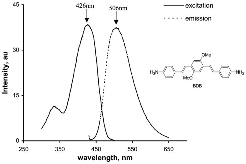
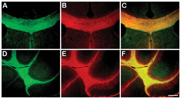
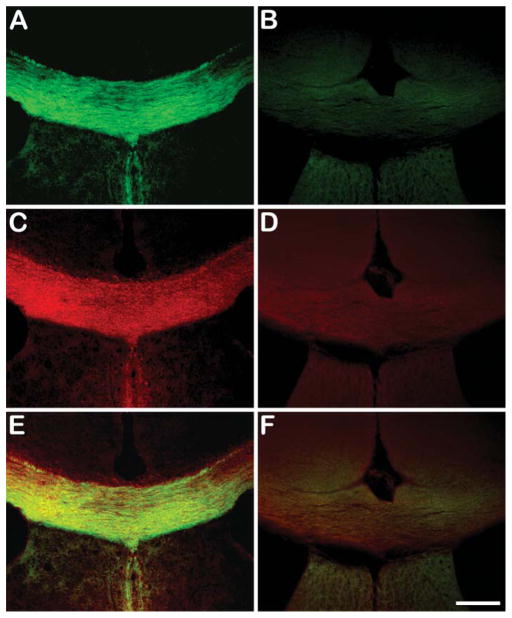
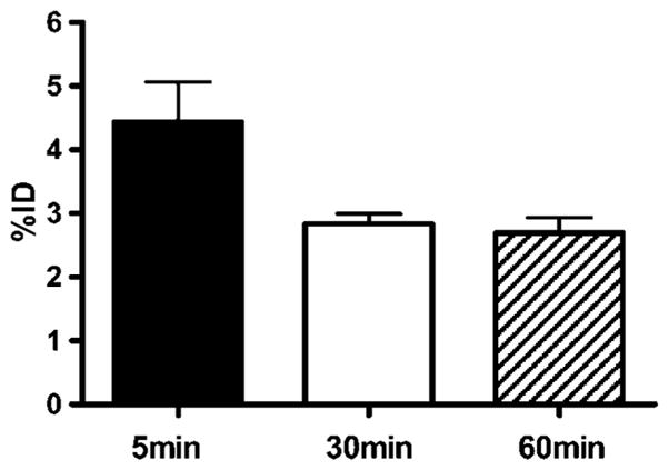
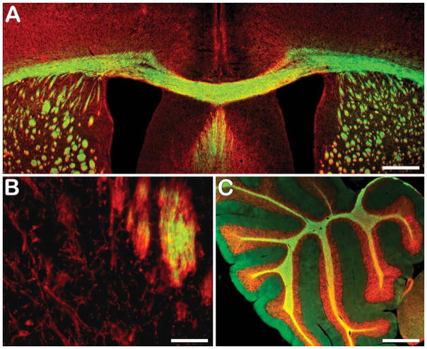
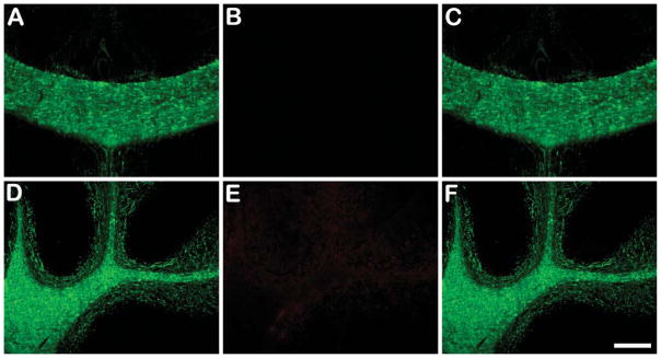
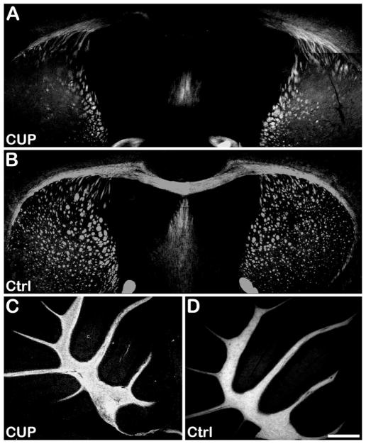
Myelin is a multilayered glial cell membrane that forms segmented sheaths around large-caliber axons of both the central nervous system (CNS) and peripheral nervous system (PNS). Myelin covering insures rapid and efficient transmission of nerve impulses. Direct visual assessment of local changes of myelin content in vivo could greatly facilitate diagnosis and therapeutic treatments of myelin-related diseases. Current histologic probes for the visualization of myelin are based on antibodies or charged histochemical reagents that do not enter the brain. We have developed a series of chemical compounds including (E,E)-1,4-bis(4'-aminostyryl)-2-dimethoxy-benzene termed BDB and the subject of this report, which readily penetrates the blood-brain barrier and selectively binds to the myelin sheath in brain. BDB selectively stains intact myelinated regions in wild-type mouse brain, which allows for delineation of cuprizone-induced demyelinating lesions in mouse brain. BDB can be injected IV into the brain and selectively detect demyelinating lesions in cuprizone-treated mice in situ. These studies justified further investigation of BDB as a potential myelin-imaging probe to monitor myelin pathology in vivo.


Figures

Figure 1
Figure 1
Excitation and emission spectra of (E,E)-1,4-bis(4′-aminostyryl)-2-dimethoxy-benzene (BDB) (1 μM in DMSO). Excitation spectra: emission at 510 nm (range 300–500 nm), bandwidth at 5 nm, scan at 120 nm/min, integration time 0.5 sec, and maximal excitation wavelength at 426 nm. Emission spectra: excitation at 426 nm (range 430–650 nm), bandwidth at 5 nm, scan at 120 nm/min, integration time of 0.5 sec, and maximal emission wavelength at 506 nm.
Figure 2
Figure 2
In vitro BDB staining of corpus callosum (green, coronal sections in A) and cerebellum (green, sagittal sections in D) in wild-type mouse brain compared with myelin basic protein (MBP) staining (red in B,E). Colocalization of corpus callosum and cerebellum with both staining are shown in C and F, respectively. Bar = 200 μm.
Figure 3
Figure 3
In vitro BDB staining of corpus callosum in wild-type mouse brain (green in A) and quaking mouse brain (B) compared with MBP staining (red in C,D). Colocalization of corpus callosum in both wild type and quaking mouse model are shown in E and F, respectively. Bar = 200 μm.
Figure 4
Figure 4
Kinetic of BDB accumulation in mice brain. BDB (300 μl of 10 mM in 10% DMSO) was injected IV into normal control mice. At the indicated times after the injection, mice were sacrificed and brain levels of BDB determined by quantitative HPLC analysis as described in Materials and Methods. Data are from n=3 mice in each group and are shown as the mean ± SD of the whole brain concentration relative to the injected dose (%ID).
Figure 5
Figure 5
Ex vivo BDB staining of myelin sheaths in the corpus callosum (green in A) and cerebellum (green in C) colocalized with MBP staining (red) in the same sections. However, ex vivo staining of oligodendrocyte cell soma present in caudate putamen with BDB showed lack of staining in the cell bodies (B), whereas immunostaining for MBP was positive, indicating that BDB preferentially stains myelinated fibers. Bars: A,C = 500 μm; B = 50 μm.
Figure 6
Figure 6
Injection of fluoromyelin in a wild-type mouse showing lack of brain entry resulting in negligible staining of myelinated corpus callosum and cerebellum in the brain. Presence of myelin sheaths in these regions was demonstrated by staining of adjacent sections with a rat anti-mouse MBP primary antibody and a FITC-conjugated goat anti-rat IgG secondary antibody. No fluoromyelin was detected in either corpus callosum (B) or cerebellum (E). In contrast, abundant MBP-positive signals were visualized in the same regions at the adjacent brain section (A,D). Merged images of corpus callosum and cerebellum from fluoromyelin and MBP staining are shown in C and F, respectively. For injection, 0.5 ml of commercial fluoromyelin was used without any dilution. Bar = 200 μm.
Figure 7
Figure 7
Ex vivo staining of corpus callosum in the cuprizone-treated mouse brain (A) and normal control mouse brain (B). Compared with the normally myelinated corpus callosum in the control mouse brain, significant demyelination was observed in the cuprizone-treated age-matched control mouse. Meanwhile, no significant demyelination was observed in cerebellum of cuprizone-treated mouse brain (C) compared with normal control mouse brain (D). CUP, cuprizone-treated; Ctrl, control. Bar = 500 μm.

References

    1. Bancroft J, Gamble M. Theory and Practice of Histological Techniques. 5. London: Churchill-Livingstone; 2002.
    1. Berube GR, Powers MM, Clark G. Iron hematoxylin chelates. I. The Weil staining bath. Stain Technol. 1965;40:53–62. - PubMed
    1. Clark SL, Ward JW. A variation of the Pal-Weigert method for staining myelin sheaths. Stain Technol. 1934;54:13–16. - PubMed
    1. Cook H. Manual of Histological Demonstration Methods. 5. London: Butterworth; 1974.
    1. Horton JC, Hocking DR. Myelin patterns in V1 and V2 of normal and monocularly enucleated monkeys. Cereb Cortex. 1997;7:166–177. - PubMed

Publication types