. 2006 May 18;34(9):2634-52.
doi: 10.1093/nar/gkl346. Print 2006.

Predicting RNA pseudoknot folding thermodynamics

Affiliations

Predicting RNA pseudoknot folding thermodynamics

Song Cao et al. Nucleic Acids Res. .

Abstract

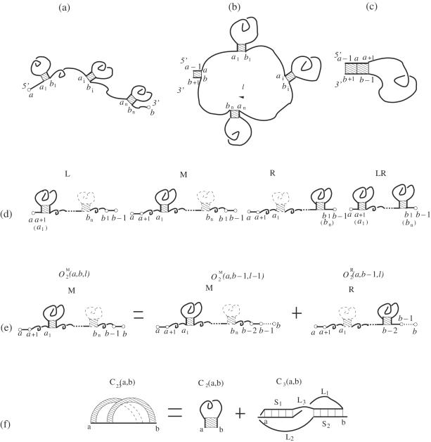
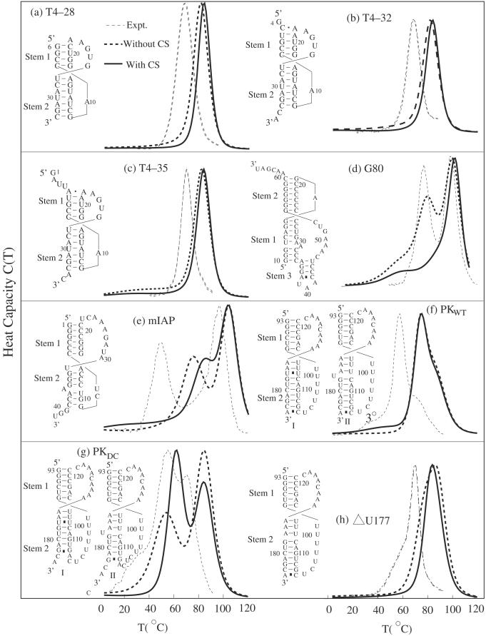
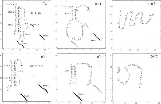
Based on the experimentally determined atomic coordinates for RNA helices and the self-avoiding walks of the P (phosphate) and C4 (carbon) atoms in the diamond lattice for the polynucleotide loop conformations, we derive a set of conformational entropy parameters for RNA pseudoknots. Based on the entropy parameters, we develop a folding thermodynamics model that enables us to compute the sequence-specific RNA pseudoknot folding free energy landscape and thermodynamics. The model is validated through extensive experimental tests both for the native structures and for the folding thermodynamics. The model predicts strong sequence-dependent helix-loop competitions in the pseudoknot stability and the resultant conformational switches between different hairpin and pseudoknot structures. For instance, for the pseudoknot domain of human telomerase RNA, a native-like and a misfolded hairpin intermediates are found to coexist on the (equilibrium) folding pathways, and the interplay between the stabilities of these intermediates causes the conformational switch that may underlie a human telomerase disease.


Figures

Figure 1
Figure 1
(a) A schematic diagram for a simple H-type pseudoknot. Loops L1 and L2 span the deep narrow (major) and the shallow wide (minor) grooves, respectively. (b) The secondary structure for the gene 32 mRNA pseudoknot of bacteriophage T2 and (c) the corresponding atomic structure for P and C4 atoms in the virtual bond representation. The atomic coordinates data are from the NMR structure (PDB ID: 2TPK). (d) The virtual bond representation for the conformation of a nucleotide strand.
Figure 2
Figure 2
(a) The C4-C4 end–end distance for the loops for different helix stem lengths. Loops L1 and L2 span across the deep and the shallow grooves of the helix stems, respectively. We obtain the C4 coordinates for short stems using the NMR determined values for the 32 mRNA pseudoknot of bacteriophage T2. For longer stems, we obtain the end–end distance from the helix coordinates generated from the virtual bond model. (b) The entropies for loops across the deep and the shallow grooves. The entropy L2, the loop across the shallow groove, shows a non-monotonic loop length-dependence.
Figure 3
Figure 3
Comparison for the calculated conformation entropy from the exact computer enumeration (Times) and the result from Equation 2 (Triangle), where L1 and L2 are the lengths (nt) for the loops across the the deep groove of S2 and the shallow groove of S1, respectively, and Ωs is the number of the pseudoknot loop conformations. The helix lengths for S1 and S2 are fixed at 5 and 7 bp, respectively (See Figure 1b).
Figure 4
Figure 4
An open conformation (a) and a closed conformation with the closing stack connected to a loop (b) or to a stack (c). The closed conformation in (b) is formed from the open conformation in (a) through the closure of the unstacked loop of length l in (b). (d) The four types of open conformation (L, M, R and LR). (e) The partition function for M-type conformations for a chain from a to b can be computed as the sum of the partition function for a shorter chain from a to b − 1. (f) A closed conformation for RNA secondary structure and pseudoknots. Here we use a polymer graph to represent C23(a,b). The straight dark lines in the polymer graph represent the nucleotide backbone. The curved links represent the base pairs between the nucleotides. The shaded region between two curved links in the polymer graph corresponds to a helix stem.
Figure 5
Figure 5
The density plots for the base pairing probabilities and the predicted stable structures for (a) TYMV and (b) TMV pseudoknots at T = 37°C with the coaxial stacking. The upper diagrams are the corresponding polymer graphs for the predicted native structures. The straight dark lines in the polymer graph represent the nucleotide backbone and the curved links represent the base pairs.
Figure 6
Figure 6
The comparison between the calculated melting curves and the experimentally measured results for eight pseudoknots sequences (a) T4–28, (b) T4–32, (c) T4–35 (24,26), (d) G80 (23), (e) mIAP (27), (f) the telomerase pseudoknot domain (PKWT), (g) (28) and (h) (29) are two mutants of the telomerase pseudoknot domain. The Y-axis is the heat capacity. The calculated melting curves have been normalized such that the theoretical and the experimental curves have the same peak value. The ion conditions are 50 mM NaCl and 1.0 mM Mg2+ for (a), (b) and (c), 1 M KCl for (d), 50 mM KCl for (e), 200 mM KCl for (f), (g) and 200 mM NaCl for (h). Because our calculated enthalpic and entropic parameters for the base stacks are from Turner's rules for 1 M NaCl salt condition, our model generally overestimates the melting temperatures. Results with and without coaxial stacking (CS) are both presented.
Figure 7
Figure 7
The density plot for the base pairing probabilities, the predicted stable structures, and the density plot for the free energy landscapes for T4–35 pseudoknot at different temperatures. In the free energy landscape F(n, nn), darker color means lower free energy. n and nn denote the numbers of the native and the non-native base pairs, respectively. At T = 70°C, the partially unfolded pseudoknot structure (Z) coexists with the hairpin structure (X).
Figure 8
Figure 8
The density plot for the base pairing probabilities and the predicted stable structures for (a) the G80 and (b) mIAP pseudoknots at different temperatures.
Figure 9
Figure 9
The density plot for the base pairing probabilities and the predicted stable structures for (a) modified telomerase pseudoknot domain (PKWT) in human and (b) its mutant (PKDC) at different temperatures.
Figure 10
Figure 10
The density plot for the base pairing probabilities and the predicted stable structures for (a) the telomerase pseudoknot domain in Tetrahymena thermophia and its mutants (b), (c), (d), (e) and (f) at different temperatures. We consider the coaxial stacking in the predictions.

References

    1. Schimmel P. RNA pseudoknots that interact with components of the translation apparatus. Cell. 1989;58:9–12. - PMC - PubMed
    1. Pleij C.W.A., Bosch L. RNA pseudoknots—structure, detection, and prediction. Meth. Enzymol. 1989;180:289–303. - PubMed
    1. Pleij C.W.A. Pseudoknots—a new motif in the RNA game. Trends Biochem. Sci. 1990;15:143–147. - PubMed
    1. Draper D.E. Pseudoknots and control of protein synthesis. Curr. Opin. Cell Biol. 1990;2:1099–1103. - PMC - PubMed
    1. Tinoco I., Jr, Puglisi J.D., Wyatt J.R. RNA Folding. In: Eckstein F., Lilley D., editors. Nucleic Acids and Molecular Biology. vol. 4. Berlin: Springer-Verlag press; 1990. pp. 205–226.

Publication types