. 2006 Aug 15;174(4):428-36.
doi: 10.1164/rccm.200503-509OC. Epub 2006 May 18.

Environmental tobacco smoke suppresses nuclear factor-kappaB signaling to increase apoptosis in infant monkey lungs

Affiliations

Environmental tobacco smoke suppresses nuclear factor-kappaB signaling to increase apoptosis in infant monkey lungs

Cai-Yun Zhong et al. Am J Respir Crit Care Med. .

Abstract

Rationale: Exposure to environmental tobacco smoke in early life has adverse effects on lung development. Apoptosis plays an essential role in development; however, the molecular mechanisms of pulmonary apoptosis induced by environmental tobacco smoke is unknown.

Objectives: To investigate the mechanistic role of nuclear factor (NF)-kappaB, a critical cell survival pathway, in the developing lungs exposed to environmental tobacco smoke.

Methods: Timed-pregnant rhesus monkeys and their offspring were exposed to filtered air or to aged and diluted sidestream cigarette smoke as a surrogate to environmental tobacco smoke (a total suspended particulate concentration of 0.99 mg/m(3) for 6 h/d, 5 d/wk) from 45-50 d gestational age to 72-77 d postnatal age (n = 4/group).

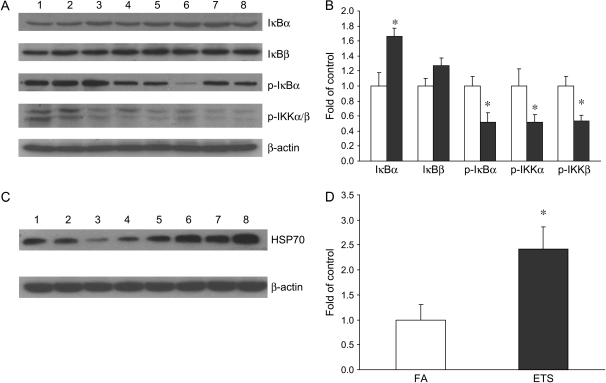
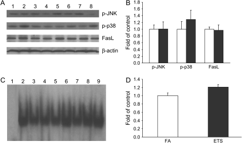
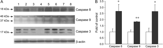
Measurements and main results: NF-kappaB-DNA binding activity, regulated anti-apoptotic genes, and apoptosis were measured in lung tissues. Exposure to environmental tobacco smoke significantly suppressed NF-kappaB activation pathway and activity. Environmental tobacco smoke further down-regulated NF-kappaB-dependent anti-apoptotic genes and induced activation of caspases, cleavage of cellular death substrates (poly(ADP)-ribose polymerase and caspase-activated DNase) and an increase in the rate of apoptosis in the lung parenchyma. No significant alterations were observed for activator protein 1, p53 or Akt activity.

Conclusions: Our results indicate that exposure to low levels of environmental tobacco smoke during a critical window of maturation in the neonatal nonhuman primate may compromise lung development with potential implications for future lung growth and function. These findings support our hypothesis that NF-kappaB plays a key role in the regulation of the apoptotic process.


Figures

<b>Figure 1.</b>
Figure 1.
Effect of environmental tobacco smoke (ETS) on NF-κB activity. (A) Electrophoretic mobility shift assay of NF-κB–DNA binding activity in the lungs of infant monkeys. Lane 1, competition assay; lanes 2–5, filtered air (FA); lanes 6–9, ETS. (B) Densitometry of NF-κB–DNA binding activity. Values are expressed as means ± SE (n = 4). Exposure to ETS significantly decreased NF-κB– DNA binding activity. *p < 0.05, compared with FA control. (C) Supershift assay of NF-κB p65, p50, and c-Rel. A supershift band for p65 as well as a weak supershift band for p50 were noted in the NF-κB–DNA complex.
<b>Figure 2.</b>
Figure 2.
Effect of ETS on NF-κB activation pathway. (A) Western blot analysis of IκBα, IκBβ, phospho-IκBα, phospho-IKKα, and phospho-IKKβ. Lanes 1–4, FA; lanes 5–8, ETS. (B) Densitometry of Western blot for IκBα, IκBβ, phospho-IκBα, phospho-IKKα, and phospho-IKKβ. There was a significant increase of IκBα, as well as significant decreases of phospho-IκBα, phospho-IKKα, and phospho-IKKβ in infant monkeys exposed to ETS (filled bars). Open bars, FA. No significant change was observed for IκBβ. (C) Western blot analysis of HSP 70. D: densitometry of Western blot for HSP 70. Values are presented as means ± SE (n = 4). There was a significant increase of HSP70 in ETS-exposed infant monkeys. *p < 0.05, compared with FA control.
<b>Figure 3.</b>
Figure 3.
Effect of ETS on the expression of Bcl-2 and XIAP mRNA. Quantitative detection of Bcl-2 and XIAP mRNA was performed using real time RT-PCR. Values for Bcl-2 and XIAP mRNA expression were normalized to the expression of GAPDH. Data are expressed as means ± SE (n = 4). Exposure to ETS (filled bars) significantly decreased the expression of Bcl-2 and XIAP mRNA. *p < 0.05, **p < 0.01, compared with FA control (open bars).
<b>Figure 4.</b>
Figure 4.
Effect of ETS on the expression of NF-κB–dependent anti-apoptotic proteins. (A) Western blot analysis of Bcl-2, Bcl-xl, TRAF 1, TRAF 2, c-IAP1, and XIAP in the lungs of infant monkeys. Lanes 1–4, FA; lanes 5–8, ETS. (B) Densitometry of Western blot. Values are presented as means ± SE (n = 4). Exposure to ETS (filled bars) significantly decreased the expression of Bcl-2, Bcl-xl, TRAF1, TRAF2, and XIAP proteins. *p < 0.05, **p < 0.01, compared with FA control (open bars).
<b>Figure 5.</b>
Figure 5.
Effect of ETS on JNK/p38/c-Jun/FasL pathway. (A) Western blot analysis of phospho-JNK, phospho-p38, and FasL. Lanes 1–4, FA; lanes 5–8, ETS. (B) Densitometry of Western blot. (C) Electrophoretic mobility shift assay of AP-1–DNA binding activity. Lane 1, competition assay; lanes 2–5, FA; lanes 6–9, ETS. (D) Densitometry of electrophoretic mobility shift assay. Values are presented as means ± SE (n = 4). No significant changes were noted for phospho-JNK, phospho-p38, FasL, or AP-1–DNA binding activity between FA control (open bars) and ETS (filled bars) groups.
<b>Figure 6.</b>
Figure 6.
Effect of ETS on p53 and Bax. (A) Western blot analysis of p53 and Bax in the lungs of infant monkeys . Lanes 1–4, FA; lanes 5–8, ETS. (B) Densitometry of Western blot. (C) Electrophoretic mobility shift assay of p53–DNA binding activity. Lane 1, competition assay; lanes 2–5, FA; lanes 6–9, ETS. (D) Densitometry of electrophoretic mobility shift assay. Values are presented as means ± SE (n = 4). No significant changes were noted for p53, Bax, or p53-DNA binding activity between FA control (open bars) and ETS (filled bars) groups.
<b>Figure 7.</b>
Figure 7.
Effects of ETS on caspase activation. (A) Western blot analysis of caspase 8, 9, and 3 in the lungs of infant monkeys. Lanes 1–4, FA; lanes 5–8, ETS. (B) Densitometry of Western blot. Values are presented as means ± SE (n = 4). There were significant increases of cleaved caspase 8, 9, and 3 in ETS-exposed infant monkeys (filled bars). *p < 0.05, **p < 0.01, compared with FA control (open bars).
<b>Figure 8.</b>
Figure 8.
Effect of ETS on cleaved PARP and CAD. (A) Western blot analysis of cleaved PARP and CAD in the nucleus of the lungs of infant monkeys. Lanes 1–4, FA; lanes 5–8, ETS. (B) Densitometry of Western blot. Values are presented as means ± SE (n = 4). The levels of cleaved PARP and CAD were significantly increased in infant monkeys exposed to ETS (filled bars). *p < 0.05, compared with FA control (open bars).
<b>Figure 9.</b>
Figure 9.
Effect of ETS on apoptosis in the lungs. TUNEL labeling of infant monkey lungs in airways (A and B) and lung parenchyma (C and D) for FA (A and C) and ETS (B and D). TUNEL-positive cells were identified as darkly stained cells, as indicated by arrows.
<b>Figure 10.</b>
Figure 10.
Apoptotic index (number of TUNEL-positive cells per total numbers of cells). Values are presented as means ± SE (n = 4). Exposure to ETS significantly increased apoptosis in the parenchyma of infant monkeys. *p < 0.05, compared with filtered air control.
<b>Figure 11.</b>
Figure 11.
Proposed model of NF-κB signaling in ETS-induced apoptotic process in the lungs of infant monkeys. Through suppression of IKK activation and IκBα degradation, as well as induction of HSP 70, ETS inhibits NF-κB activity and downregulates the expression of NF-κB–dependent anti-apoptotic genes, including members of Bcl-2, TRAFs, and c-IAPs, resulting in activation of initiator caspases and effector caspase. The effector caspase 3 cleaves the key death substrates such as PARP and ICAD/CAD, leading to the apoptotic process.

References

    1. World Health Organization Division of Noncommunicabe Disease Tobacco Free Initiative. International consultation on environmental tobacco smoke (ETS) and child health. Consultation Report, Geneva, 1999. http://www.who.int/toh.
    1. Gergen PJ, Fowler JA, Maurer KR, Davis WW, Overpeck MD. The burden of environmental tobacco smoke exposure on the respiratory health of children 2 months through 5 years of age in the United States: Third National Health and Nutrition Examination Survey, 1988 to 1994. Pediatrics 1998;101:8–13. - PubMed
    1. Finkelstein JN, Johnston CJ. Enhanced sensitivity of the postnatal lung to environmental insults and oxidant stress. Pediatrics 2004;113:1092–1096. - PubMed
    1. Pinkerton KE, Joad JP. The mammalian respiratory system and critical windows of exposure for children's health. Environ Health Perspect 2000;108:457–462. - PMC - PubMed
    1. DiFranza JR, Aligne CA, Weitzman M. Prenatal and postnatal environmental tobacco smoke exposure and children's health. Pediatrics 2004;113:1007–1015. - PubMed

Publication types