. 2006 Jun 21;25(12):2735-45.
doi: 10.1038/sj.emboj.7601153. Epub 2006 May 18.

C-terminal-binding protein directly activates and represses Wnt transcriptional targets in Drosophila

Affiliations

C-terminal-binding protein directly activates and represses Wnt transcriptional targets in Drosophila

Ming Fang et al. EMBO J. .

Abstract

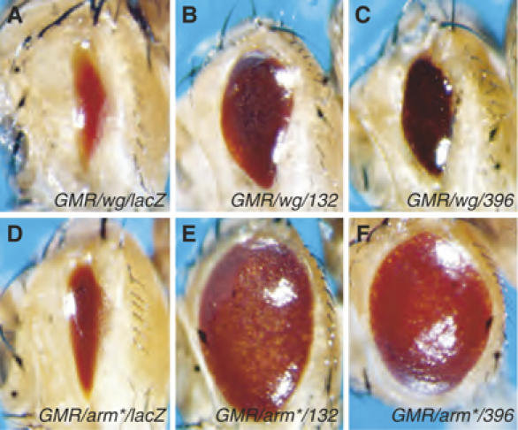
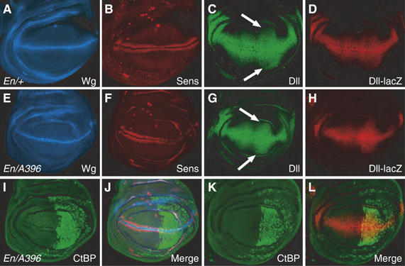
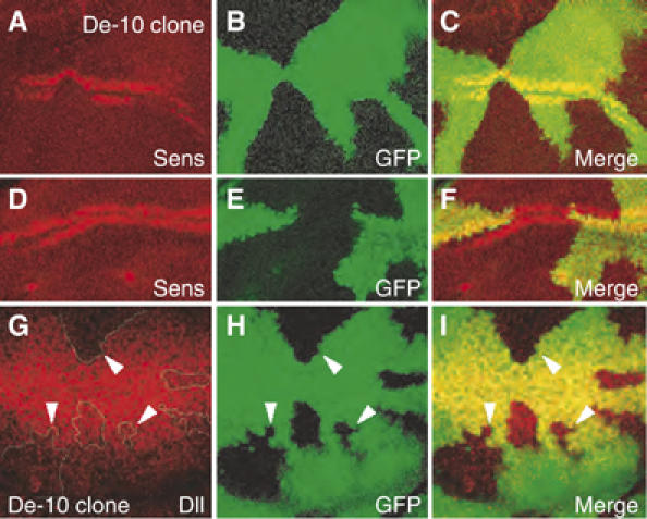
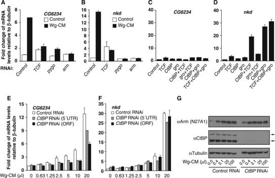
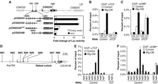
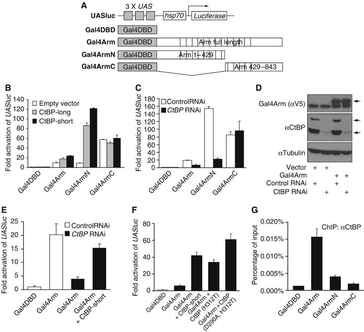
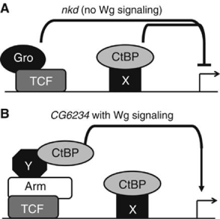
Regulation of Wnt transcriptional targets is thought to occur by a transcriptional switch. In the absence of Wnt signaling, sequence-specific DNA-binding proteins of the TCF family repress Wnt target genes. Upon Wnt stimulation, stabilized beta-catenin binds to TCFs, converting them into transcriptional activators. C-terminal-binding protein (CtBP) is a transcriptional corepressor that has been reported to inhibit Wnt signaling by binding to TCFs or by preventing beta-catenin from binding to TCF. Here, we show that CtBP is also required for the activation of some Wnt targets in Drosophila. CtBP is recruited to Wnt-regulated enhancers in a Wnt-dependent manner, where it augments Armadillo (the fly beta-catenin) transcriptional activation. We also found that CtBP is required for repression of a subset of Wnt targets in the absence of Wnt stimulation, but in a manner distinct from previously reported mechanisms. CtBP binds to Wnt-regulated enhancers in a TCF-independent manner and represses target genes in parallel with TCF. Our data indicate dual roles for CtBP as a gene-specific activator and repressor of Wnt target gene transcription.


Figures

Figure 1
Figure 1
Two GSV insertions in the CtBP locus suppress Wg and Arm-dependent signaling in the eye. Micrographs of adult fly heads containing P[GMR-Gal4] and P[UAS-wg] (A–C) or P[GMR-Arm*] (D–F) and the following transposons: (A, D) P[UAS-lacZ], (B, E) P[GSV]A132 or (C, F) P[GSV]A396. Expression of wg via the GMR promoter produces an eye that is severely reduced in size (A), and this phenotype is suppressed by A132 or A396 (B, C). Expression of an activated form of Arm (Arm*) also causes eye size reduction (D) that is dramatically suppressed by A132 or A396 (E, F). Each transgene is present in one copy/fly and flies were reared at 25°C.
Figure 2
Figure 2
CtBP overexpression can activate and repress Wg targets in the wing imaginal discs. Confocal images of wing imaginal discs from late third instar larva. (A, B) P[En-Gal4]/+ disc immunostained for Wg (blue) and Sen (red) displaying the wild-type expression pattern. (C, D) P[En-Gal4]/P[Dll-lacZ] discs stained for Dll (green) and lacZ (red) showing the normal broad expression surrounding the D/V boundary. (E, F, I, J) P[En-Gal4]/P[GSV]A396 disc, where CtBP (green) is overexpressed in the posterior compartment. Wg expression (blue) is unaffected, while Sens (red) is sharply reduced in the CtBP-expressing domain. (G) P[En-Gal4]/P[GSV]A396 disc stained for Dll (green), displaying a subtle but reproducible expansion of Dll expression in the posterior compartment (compare arrows in panel G with those in panel C). (H, K, L) P[En-Gal4] P[GSV]A396 P[Dll-lacZ] disc stained for CtBP (green) and lacZ (red), exhibiting a significantly wider Dll-lacZ expression domain in the posterior compartment.
Figure 3
Figure 3
Loss of endogenous CtBP results in a reduction in activation of Wg targets in the wing imaginal discs. CtBP activity was removed by creating mitotic clones of CtBP87De-10 (De-10), a strong CtBP allele. Clones are marked by the absence of GFP (green). Clones displayed a highly penetrant (100%, n=8) loss of Sens expression at ∼12–15 h before pupariation (A–C), which was not observed in clones from discs that were a few hours prior to pupariation (D–F). A reduction in Dll expression (90% penetrance, n=30) was observed in late third instar discs (G–I), which was more pronounced in clones further from the D/V boundary (white arrowheads).
Figure 4
Figure 4
CtBP represses as well as activates endogenous Wg targets in Kc cells. (A, B) Kc cells were treated with control dsRNA or sequences corresponding to TCF, pygo and arm for 4 days before the addition of control media or Wg-CM for 4 h. Transcript levels of CG6234 (A) and nkd (B) were measured by quantitative RT–PCR as described in Materials and methods. Results were normalized to β-tubulin56D expression. The RNAi efficiencies for each gene were monitored by Western analysis to ensure that the corresponding protein levels were significantly reduced (data not shown). The induction of CG6234 and nkd expression by Wg-CM was severely reduced by TCF, Pygo or Arm depletion. (C, D) Synergistic derepression of nkd but not CG6234 by CtBP RNAi combined with the RNAi of TCF or gro. Kc cells were treated with the indicated dsRNAs for 4 days before harvesting and expression analysis as described above. (E, F) Cells were incubated with control dsRNA or duplexes specific for the 5′UTR or ORF of CtBP for 4 days before stimulation with increasing amounts of Wg-CM for 4 h before analysis of CG6234 (E) and nkd (F) expression as described above. Each bar represents the mean of duplicate cultures and duplicate transcript determinations, with the lines indicating the standard error. All experiments have been performed at least three separate times and representative experiment is shown. (G) Western analysis with anti-Arm and anti-CtBP antibody in control or CtBP RNAi-treated cells with increasing amounts of Wg-CM. CtBP RNAi severely affected CtBP expression (the two arrows indicate the short and long CtBP isoforms) but had no effect on Arm stabilization by Wg-CM. αTubulin levels are used as a loading control. The gels shown are representative of three separate experiments.
Figure 5
Figure 5
CtBP binding to WREs is activated by Wg signaling, but is TCF-independent in the absence of Wg. (A) Schematic diagram of the CG6234 locus showing the location of the predicted TCF sites (C#1 and 2) and a coding region control site (C#0) used for ChIP analysis. pCG6234 is a 1.7 kb fragment containing the predicted TCF sites (vertical lines) fused to a hsp70 core promoter and luciferase. This reporter construct, and one containing the 5′ 1133 bp of the fragment (pCG6234A) are activated by co-transfection of Arm*. This activation is abolished by mutations of all the potential TCF sites (pCG6234Amut). Each bar is the mean of duplicate transfections where luciferase activity was determined in duplicate, with the standard error indicated by the lines. The data shown is a representative example from three separate experiments. (B) TCF binding to C#1 and the C#0 control, as assayed by ChIP in cells treated with control or TCF dsRNA and stimulated with control or Wg-CM for 4 h. Wg stimulation increases TCF binding, and little signal is observed in TCF-depleted cells. (C) Cells treated as in (B) analyzed for CtBP binding. Wg stimulation increased the CtBP ChIP signal on the WRE, and this increase is abolished in TCF-depleted cells. However, CtBP binding in the absence of Wg is TCF-independent. (D) nkd locus with the predicted TCF site clusters (N#1–7) and a coding region control site (N#0) for ChIP analysis. (E, F) ChIP analysis shows overlapping binding of TCF (E) and CtBP (F) to the nkd control region. Binding of both proteins is increased by a 4 h treatment of Wg-CM, and CtBP Wg-dependent recruitment is blocked in TCF-depleted cells. As with CG6234, CtBP binding in the absence of Wg is TCF-independent. For both loci, in the absence of Wg signaling, there is a reproducible increase in the CtBP ChIP signal in TCF-depleted cell extracts. It should also be noted that relative strength of the ChIP signal for N#4 and N#5 sites in nkd varies from experiment to experiment for both TCF and CtBP. The proximity of these primers (352 bp) is less than the resolution of ChIP (based on the size of the sonicated fragments). However, these primer sets always give higher signals than primers corresponding to other regions of the nkd locus. The bars for each ChIP signal are the mean of duplicate precipitations, with duplicate Q-PCR reactions. Standard errors are indicated by the lines and each experiment has been carried out at least three times with similar results.
Figure 6
Figure 6
CtBP is required for Arm-dependent transcription activity. (A) Schematic diagram of the UAS reporter and the Gal4 expression vectors used in the following experiments. (B) Overexpression of both short and long forms of CtBP (500 ng/well) enhances the transcription activities of Gal4Arm (50 ng/well) and Gal4ArmN but not Gal4ArmC (each 20 ng/well) on the UASluc reporter in Kc cells. Cells were transfected and luciferase and β-galactosidase activities assayed as described in Materials and methods. (C) CtBP RNAi using a dsRNA corresponding to the 5′UTR diminishes the transcription activities of Gal4Arm and Gal4ArmN but not Gal4ArmC (all 100 ng/well) on UASluc reporter. (D) Western blot analysis shows the expression levels of Gal4Arm (V5 tagged) and short and long isoforms of CtBP in cells with RNAi and transfection as indicated. CtBP RNAi severely reduced the amount of endogenous CtBP, but had no effect on transfected Gal4Arm (the band below Gal4Arm is non-specific). (E) The effect of CtBP RNAi (5′UTR) can be rescued by a CtBP transgene containing a heterologous 5′UTR. (F) Mutations of conserved catalytic residues (D290A and H312T) in CtBP had no effect on enhancement on UASluc reporter activity with Gal4Arm (5 ng/well). (G) CtBP ChIP in cells co-transfected with UASluc (1 ng/well) and Gal4DBD, Gal4Arm, Gal4ArmN or Gal4ArmC constructs (500 ng/well each) shows enhanced occupancy of CtBP on the UAS sites of UASluc in cells containing Gal4Arm and Gal4ArmN compared with Gal4DBD. Gal4ArmC did not show a significant difference with Gal4DBD in several experiments. Each bar is the mean of duplicate transfections and duplicate luciferase or Q-PCR reactions, with the lines indicating the standard errors. Each experiment was performed at least three times and similar results were obtained in each experiment.
Figure 7
Figure 7
Model depicting CtBP functions in the absence or presence of Wg signaling. (A) In the absence of Wg signaling, CtBP (presumably recruited to the WRE by an unknown protein) acts in parallel to TCF/Gro to repress nkd gene expression. (B) Upon Wg stimulation, CtBP is recruited by Arm and other factors to the TCF binding sites, where it contributes to activation of targets such as CG6234.

References

    1. Barker N, Hurlstone A, Musisi H, Miles A, Bienz M, Clevers H (2001) The chromatin remodelling factor Brg-1 interacts with beta-catenin to promote target gene activation. EMBO J 20: 4935–4943 - PMC - PubMed
    1. Barolo S, Posakony JW (2002) Three habits of highly effective signaling pathways: principles of transcriptional control by developmental cell signaling. Genes Dev 16: 1167–1181 - PubMed
    1. Bauer A, Huber O, Usseglio F, Rothbacher U, Aragnol D, Kemler R, Pradel J (2000) Pontin 52 and reptin 52 function as antagonistic regulators of beta-catenin signalling activity. EMBO J 19: 6121–6130 - PMC - PubMed
    1. Brannon M, Brown JD, Bates R, Kimelman D, Moon RT (1999) XCtBP is a XTcf-3 co-repressor with roles throughout Xenopus development. Development 126: 3159–3170 - PubMed
    1. Cadigan KM (2002) Regulating morphogen gradients in the Drosophila wing. Semin Cell Dev Biol 13: 83–90 - PubMed

Publication types

MeSH terms