. 2006 Jun 7;25(11):2358-67.
doi: 10.1038/sj.emboj.7601149. Epub 2006 May 18.

UBP43 is a novel regulator of interferon signaling independent of its ISG15 isopeptidase activity

Affiliations

UBP43 is a novel regulator of interferon signaling independent of its ISG15 isopeptidase activity

Oxana A Malakhova et al. EMBO J. .

Abstract

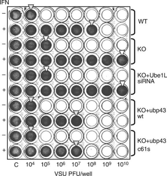
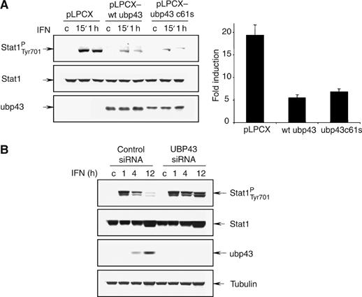
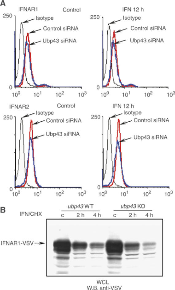
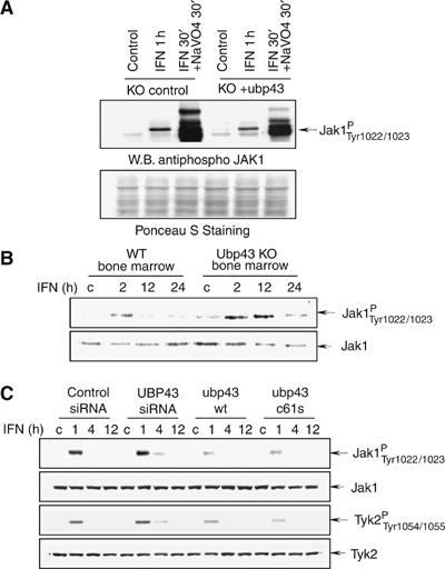
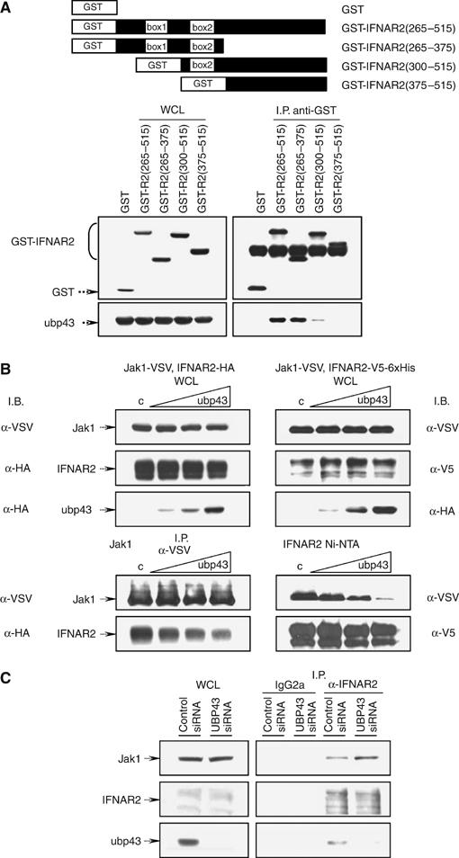
Interferons (IFNs) regulate diverse cellular functions through activation of the Janus kinase-signal transducer and activator of transcription (JAK-STAT) pathway. Lack of Ubp43, an IFN-inducible ISG15 deconjugating enzyme, leads to IFN hypersensitivity in ubp43-/- mice, suggesting an important function of Ubp43 in downregulation of IFN responses. Here, we show that Ubp43 negatively regulates IFN signaling independent of its isopeptidase activity towards ISG15. Ubp43 functions specifically for type I IFN signaling by downregulating the JAK-STAT pathway at the level of the IFN receptor. Using molecular, biochemical, and genetic approaches, we demonstrate that Ubp43 specifically binds to the IFNAR2 receptor subunit and inhibits the activity of receptor-associated JAK1 by blocking the interaction between JAK and the IFN receptor. These data implicate Ubp43 as a novel in vivo inhibitor of signal transduction pathways that are specifically triggered by type I IFN.


Figures

Figure 1
Figure 1
Negative regulation of IFN signaling by Ubp43 is independent of its enzymatic activity. (A) Total protein extracts were prepared from ubp43+/+ and ubp43−/− MEFs or ubp43−/− MEFs stably expressing Ube1L siRNA, wt murine Ubp43, or Ubp43C61S after mIFN-β (1000 U/ml) treatment for 0′, 30′, and 10 h and analyzed by Western blotting with the respective antibodies. (B) 293T cells were transiently transfected with vector-control (lane 1) or plasmids containing 6xHis-ISG15, HA-Ube1L, and Flag-Ubc8 in the absence (lane 2) or the presence of wt (lane 3) or mutant (c61s) Ubp43-V5 (lane 4). The level of ISG15 conjugation was determined by Western blotting with anti-mISG15 antibodies. Blots were stripped and reprobed with anti-HA, anti-Flag, or anti-V5 antibodies to ensure equal levels of protein expression.
Figure 2
Figure 2
VSV protection assay. ubp43+/+, ubp43−/−, ubp43−/− MEFs with stable expression of Ube1L siRNA, wt Ubp43, or Ubp43C61S were left untreated or treated with 1000 U/ml of mIFN-β for 24 h, followed by mock or VSV infection ranging from 104 to 1010 PFU/well for additional 24 h. Cell viability was assessed by crystal violet staining. The arrows indicate the level of protection.
Figure 3
Figure 3
Ectopic expression of Ubp43 blocks STAT1 phosphorylation and IFN-mediated gene induction. (A) STAT1-deficient U3A stable cell lines expressing vector-control, wt mUbp43, or mUbp43C61S were transiently co-transfected by STAT1 and ISRE-driven luciferase reporter plasmid. At 24 h post-transfection, cells were either left untreated or treated with hIFN-α for 15′, 1 h, and 16 h. Level of STAT1 phosphorylation and expression was assessed by Western blotting with the respective antibodies (left). Luciferase activities were measured, normalized, and presented as fold increase of relative luciferase activity in IFN treated cells (at 16 h point) over the untreated controls (right). The error bars indicate the s.d. of the mean. (B) KT-1 cells were stably transfected with control siRNA, hUBP43-specific siRNA. After 1 week of drug selection, cells were either left untreated or treated with hIFN-α for 1, 4 or 12 h respectively. STAT1 phosphorylation and expression was assessed by Western blotting with the respective antibodies. Specific inhibition of endogenous hUBP43 by siRNA in the respective stable lines was confirmed by Western blotting with anti-hUBP43-specific antibodies.
Figure 4
Figure 4
IFN receptor cell surface expression and degradation are not altered in Ubp43 deficient cells. (A) The steady-state or IFN-induced level of the endogenous IFNAR1 (upper left panels) and IFNAR2 (lower left panels) chains was determined by flow cytometry analysis of KT-1 cells, expressing control or UBP43-siRNA by using polyclonal anti-IFNAR1 and monoclonal MMHAR-2 antibodies, respectively. Graphs, corresponding to the isotype-matched control or IFNAR-specific staining in KT-1 control and UBP43-siRNA cells are marked by arrows. (B) Protein extracts were prepared from ubp43+/+ and ubp43−/− MEFs with stable expression of VSV-tagged mIFNAR1 that were either left untreated or treated with IFN and cycloheximide (CHX, to inhibit the protein synthesis) for 2 and 4 h respectively. The rate of IFNAR1 degradation was assessed by Western blotting.
Figure 5
Figure 5
Ubp43 inhibits the activation of JAK-kinases. (A) Whole-cell lysates (WCL) were prepared from ubp43−/− MEFs or ubp43−/− MEFs reconstituted with wt mUbp43 after treatment with mIFN-β alone or mIFN-β+sodium orthovanadate for the indicated period of time. Level of JAK1 phosphorylation was determined by Western blotting using phospho-specific antibodies against JAK1 (pYpY1022/1023) (upper panel). After SDS–PAGE membrane was stained with Ponceau S solution to assure equal protein loading (lower panel). (B) Bone marrow cells were prepared from ubp43+/+ or ubp43−/− mice and incubated with 100 U/ml of mIFN-β for various periods of time as indicated in the figure. JAK1 was immunoprecipitated from WCL and subjected to an in vitro kinase assay, followed by immunoblotting with anti-phosphospecific JAK1 antibodies. Blots were stripped and re-probed with anti-JAK1 antibodies. (C) KT-1 cells stably expressing control siRNA, human UBP43-specific siRNA, plasmids encoding wt Ubp43 or Ubp43C61S mutant protein were stimulated with hIFN-α (1000 U/ml) for the indicated periods of time. WCL were subjected to immunoblotting with antiphosphospecific JAK1 and TYK2 antibodies. Blots were stripped and re-probed with anti-JAK1 and TYK2 antibodies respectively to assure equal protein loading.
Figure 6
Figure 6
Ubp43 interacts with IFNAR2 receptor subunit. (A) 293T cells were transiently transfected with Flag-IFNAR1, HA-IFNAR2, or Flag-IFNGR1 and either GST control or GST-Ubp43. Reciprocal immunoprecipitations (I.P.) were performed using anti-Flag/HA or anti-GST antibodies. Whole-cell lysates (WCL) or immunoprecipitated complexes were subjected to immunoblotting with anti-HA antibodies (top middle panel), anti-Flag (top left & right panels) or anti-GST (bottom panel) antibodies, respectively. (B) Transient co-transfections of 293T cells were performed using HA-tagged IFNAR2 and various deletion mutants of Ubp43 (a.a. positions are indicated in the figure). Ubp43 deletion constructs used for this study are schematically represented in the top panel. WCL or immunoprecipitated (anti-HA) complexes were subjected to immunoblotting with anti-HA antibodies (middle panel) or anti-GST (bottom panel) antibodies, respectively. (C) wt Ubp43 and Ubp43 mutants: D331K340-AA, R350R352R354-AAA, K364-A (positions of a.a. substitutions are graphically shown on the top panel of the figure) were co-expressed with GST-IFNAR2 (a.a. 265–515) in 293T cells. I.P. were performed using anti-HA antibodies followed by immunoblotting with anti-HA antibodies (top) to verify equal loading and anti-GST-antibodies (bottom). (D) U3A cells were transiently co-transfected by the combination of ISRE-driven luciferase reporter plasmid, STAT1, and either vector-control, wt Ubp43, or Ubp43 mutants: D331K340-AA (DK), R350R352R354-AAA (RRR) and K364-A (K). At 24 h post-transfection, cells were either left untreated or treated with hIFN-α for 24 h. Luciferase activities were measured, normalized, and presented as fold increase of relative luciferase activity in IFN treated cells over the untreated controls (average of 4 independent experiments). The error bars indicate the s.d. of the mean.
Figure 7
Figure 7
Ubp43 competes with JAK1 for receptor binding. (A) 293T cells were transiently co-transfected with HA-Ubp43 and GST or GST-IFNAR2 truncation constructs schematically presented in the top panel of the figure. Whole cell lysate (WCL) (left panels) or immunoprecipitated complexes (right panels) were subjected to immunoblotting against GST or HA-tag, respectively. (B) 293T cells were transiently co-transfected with JAK1-VSV and IFNAR2-HA (left) or JAK1-VSV and IFNAR2-V5-6xHis (right) in the absence or presence of increasing concentration of HA-Ubp43 followed by immunoprecipitation using antibodies against VSV-tagged JAK1 (left, bottom panels) or Ni-NTA purification of 6xHis-tagged IFNAR2 (right, bottom panels). WCL (top panels) or immunoprecipitated complexes (bottom panels) were subjected to immunoblotting with antibodies indicated in the figure. (C) Protein extracts from stable KT-1 transfectants expressing either control siRNA or UBP43 specific siRNA and treated with hIFN-α for 4 h were used for the immunoprecipitations with control IgG2a antibodies or with anti-IFNAR2 antibodies. WCL and immunoprecipitates were subjected to immunoblotting with anti-JAK1 (top panel), anti-IFNAR2 (middle panel), and anti-Ubp43 antibodies (bottom panel).

References

    1. Aaronson DS, Horvath CM (2002) A road map for those who don't know JAK–STAT. Science 296: 1653–1655 - PubMed
    1. Basu L, Yang CH, Murti A, Garcia JV, Croze E, Constantinescu SN, Mullersman JE, Pfeffer LM (1998) The antiviral action of interferon is potentiated by removal of the conserved IRTAM domain of the IFNAR1 chain of the interferon alpha/beta receptor: effects on JAK–STAT activation and receptor down-regulation. Virology 242: 14–21 - PubMed
    1. Biron CA (2001) Interferons alpha and beta as immune regulators—a new look. Immunity 14: 661–664 - PubMed
    1. Chawla-Sarkar M, Lindner DJ, Liu YF, Williams BR, Sen GC, Silverman RH, Borden EC (2003) Apoptosis and interferons: role of interferon-stimulated genes as mediators of apoptosis. Apoptosis 8: 237–249 - PubMed
    1. Constantinescu SN, Croze E, Wang C, Murti A, Basu L, Mullersman JE, Pfeffer LM (1994) Role of interferon-alpha/beta receptor chain-1 in the structure and transmembrane signaling of the interferon-alpha/beta receptor complex. Proc Natl Acad Sci USA 91: 9602–9606 - PMC - PubMed

Publication types

MeSH terms