. 2006 Jun 7;25(11):2315-25.
doi: 10.1038/sj.emboj.7601145. Epub 2006 May 18.

Crystal structure of CHP2 complexed with NHE1-cytosolic region and an implication for pH regulation

Affiliations

Crystal structure of CHP2 complexed with NHE1-cytosolic region and an implication for pH regulation

Youssef Ben Ammar et al. EMBO J. .

Abstract

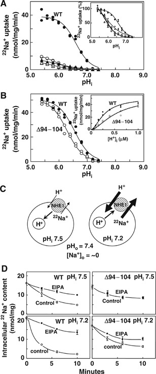
The plasma membrane Na+/H+ exchangers (NHE) require calcineurin B homologous protein (CHP) as an obligatory binding partner for ion transport. Here, we report the first crystal structure of CHP (CHP2 isoform) in complex with its binding domain in NHE1. We show that the cytoplasmic alpha-helix of NHE1 is inserted into the hydrophobic cleft formed by N- and C-lobes of CHP2 and that the size and shape of this crevice together with hydrogen bond formation at multiple positions assure a high degree of specificity for interaction with NHE members. Structure-based mutagenesis revealed the importance of hydrophobic interactions between CHP/NHE1 for the function of NHE1. Furthermore, the crystal structure shows the existence of a protruding CHP-unique region, and deletion of this region in CHP2 inhibited the NHE1 activity by inducing the acidic shift of intracellular pH dependence, while preserving interaction with NHE1. These findings suggest that CHP serves as an obligatory subunit that is required both for supporting the basic activity and regulating the pH-sensing of NHE1 via interactions between distinct parts of these proteins.


Figures

Figure 1
Figure 1
Sequence alignments of CHP and its binding domain in NHE isoforms. (A) Membrane topology model of NHE1. NHE1 consists of the N-terminal transporter domain with 12 transmembrane domains (TM) and a long C-terminal regulatory domain. Six extracellular (EL) and five intracellular loops (IL) are mapped. CHP binds to the juxtamembrane cytoplasmic domain. Arg440 was identified as an important residue for NHE1 regulation. (B) Amino-acid sequences for human NHE1–NHE5 (Genbank Accession Numbers: NM_003047, NM_003048, NM_004174, NM_177084, and NM_004594, respectively) are aligned together with sequence of CNB-binding region in CNA (Q08209). Hydrophobic residues involved in interaction with CHP or CNB are highlighted in green. Residues in NHE1 forming hydrogen bonds with CHP2 are shown in red. (C) Amino-acid sequences of CHP1/2 and CNB (Q99653, Q9D869, and P63098). Secondary structural elements are based on the structure of CHP2/NHE1-peptide complex. Loops of four EF-hands are shown by green boxes, but EF1 and EF2 are ancestral sites that do not coordinate Ca2+. Hydrophobic residues involved in interaction with NHE1 or CNA are highlighted in blue and red, respectively. Central CHP-unique region is boxed. Residues in CHP2 forming hydrogen bonds with NHE1 are shown in red.
Figure 2
Figure 2
Stereo view of the CHP2/NHE1-peptide complex showing the overall structure. N- and C-lobes of CHP2 are colored red and blue, respectively. The NHE1 peptide is colored green. Pink spheres represent the two yttrium ions coordinated by EF3 and EF4.
Figure 3
Figure 3
Target specificity of hydrophobic cleft. (A–C) Surface features of CHP2, CNB, and KChP1 are presented together with α-helices of target peptides, respectively. In CHP2, front (upper) and side (lower) views are shown. The N- and C-lobes are colored light blue and light green, while the CHP-unique region is colored red. (D, E) Surface view of NHE1 (left) and CNA (right) peptides, respectively. The upper panels indicate the side facing outside the cleft and the lower panels represent the side facing the cleft. Hydrophobic residues are colored yellow.
Figure 4
Figure 4
Closeup view showing the interaction between CHP2 and NHE1-peptide. (A) The NHE1-peptide backbone is shown in magenta, while hydrophobic side chains are shown in yellow. The hydrophobic pocket surrounding Leu527 is marked in red. His523 of NHE1 and Tyr123 of CHP2 are marked in blue. (B) Closeup view showing interaction between the N-lobe of CHP2 and the C-terminus of NHE1-peptide. (C) Ile534 is accommodated into the hydrophobic pocket produced by residues mainly in the N-lobe of CHP2.
Figure 5
Figure 5
Effects of mutations on the interaction between NHE1 and CHP2 in cells. (A) Low magnification confocal images of cells coexpressing GFP-tagged CHP2 and the wild-type (left) or I537K (right) mutant exchangers. Inset shows the intensity profile of GFP fluorescence along the dotted line in a marked cell. In most cells expressing the wild-type NHE1 but not I537K, strong fluorescent signals were detected at the cell edge. (B) Subcellular localization of CHP2 expressed in cells. GFP-tagged CHP2 was coexpressed in cells stably expressing the wild-type or mutant NHE1 variants and GFP-fluorescence was observed by confocal microscopy. (C) Subcellular localization of mutant CHP2. GFP-tagged CHP2 mutants were expressed in cells stably expressing the wild-type NHE1. Eleven residues from Glu94 to Lys104 of CHP2 were deleted in Δ94–104, while the 10 N-terminal residues from Met1 to Val10 were deleted in ΔN10. For one control experiment, GFP-tagged human CNB was expressed in NHE1-transfectants. (D) Summary data for membrane localization of GFP-tagged CHP2. Intensity profile analysis was performed on confocal images as shown in (A). The number of cells with strong fluorescence signal at the cell edge (at least three times more than the average of fluorescence in the internal cell region) was counted. Data are expressed as the mean±s.d. from 6–8 images (total cell number analyzed, 99–341).
Figure 6
Figure 6
Effect of mutations on the exchange activity. (A), pHi-dependence of EIPA-sensitive 22Na+-uptake in cells expressing wild-type NHE1 (•) or CHP binding-defective mutants, I534D (○), I534K (▴) and I537K (▵). pHi was clamped at various values with K+/nigericin. Data were fitted to Hill equations with the kinetic parameters shown in Supplementary Table I and plotted after normalization by the maximal activity at pHi=5.4 (inset). (B) pHi-dependence of EIPA-sensitive 22Na+-uptake in cells co-expressing wild-type NHE1 and GFP-tagged CHP2 (•) or deletion mutant Δ94–104 of CHP2 (○). Data were fitted to Hill equations with the kinetic parameters shown in Supplementary Table I. The 22Na+-uptake activity was also plotted against intracellular H+ concentration up to 1 μM (inset). (C) Schematic drawing of 22Na+-efflux protocol. The efflux experiment was done in 22Na+-loaded, pHi-clamped cells at extracellular pH 7.4. At lower pHi 22Na+ efflux would be accelerated by H+ binding to the regulatory site, while at high pHi it would be inhibited by H+-release from the regulatory site. (D) Time courses of 22Na+ efflux. Cells were loaded with 22Na+ and at the same time pHi-clamped at pHi 7.5 or 7.2. After removal of the radioactive solution, cells were exposed to the nonradioactive solutions with or without 0.1 mM EIPA. Data are expressed as the mean±s.d. of three determinations. Error bars are sometimes smaller than symbol sizes.

References

    1. Aharonovitz O, Zaun HC, Balla T, York JD, Orlowski J, Grinstein S (2000) Intracellular pH regulation by Na(+)/H(+) exchange requires phosphatidylinositol 4,5-bisphosphate. J Cell Biol 150: 213–224 - PMC - PubMed
    1. Ames JB, Ishima R, Tanaka T, Gordon JI, Stryer L, Ikura M (1997) Molecular mechanics of calcium-myristoyl switches. Nature 389: 198–202 - PubMed
    1. An WF, Bowlby MR, Betty M, Cao J, Ling HP, Mendoza G, Hinson JW, Mattsson KI, Strassle BW, Trimmer JS, Rhodes KJ (2000) Modulation of A-type potassium channels by a family of calcium sensors. Nature 403: 553–556 - PubMed
    1. Aronson PS, Nee J, Suhm MA (1982) Modifier role of internal H+ in activating the Na+-H+ exchanger in renal microvillus membrane vesicles. Nature 299: 161–163 - PubMed
    1. Barroso MR, Bernd KK, DeWitt ND, Chang A, Mills K, Sztul ES (1996) A novel Ca2+-binding protein, p22, is required for constitutive membrane traffic. J Biol Chem 271: 10183–10187 - PubMed

Publication types

MeSH terms