. 2006 May;2(5):e46.
doi: 10.1371/journal.ppat.0020046. Epub 2006 May 19.

Legionella pneumophila exploits PI(4)P to anchor secreted effector proteins to the replicative vacuole

Affiliations

Legionella pneumophila exploits PI(4)P to anchor secreted effector proteins to the replicative vacuole

Stefan S Weber et al. PLoS Pathog. 2006 May.

Abstract

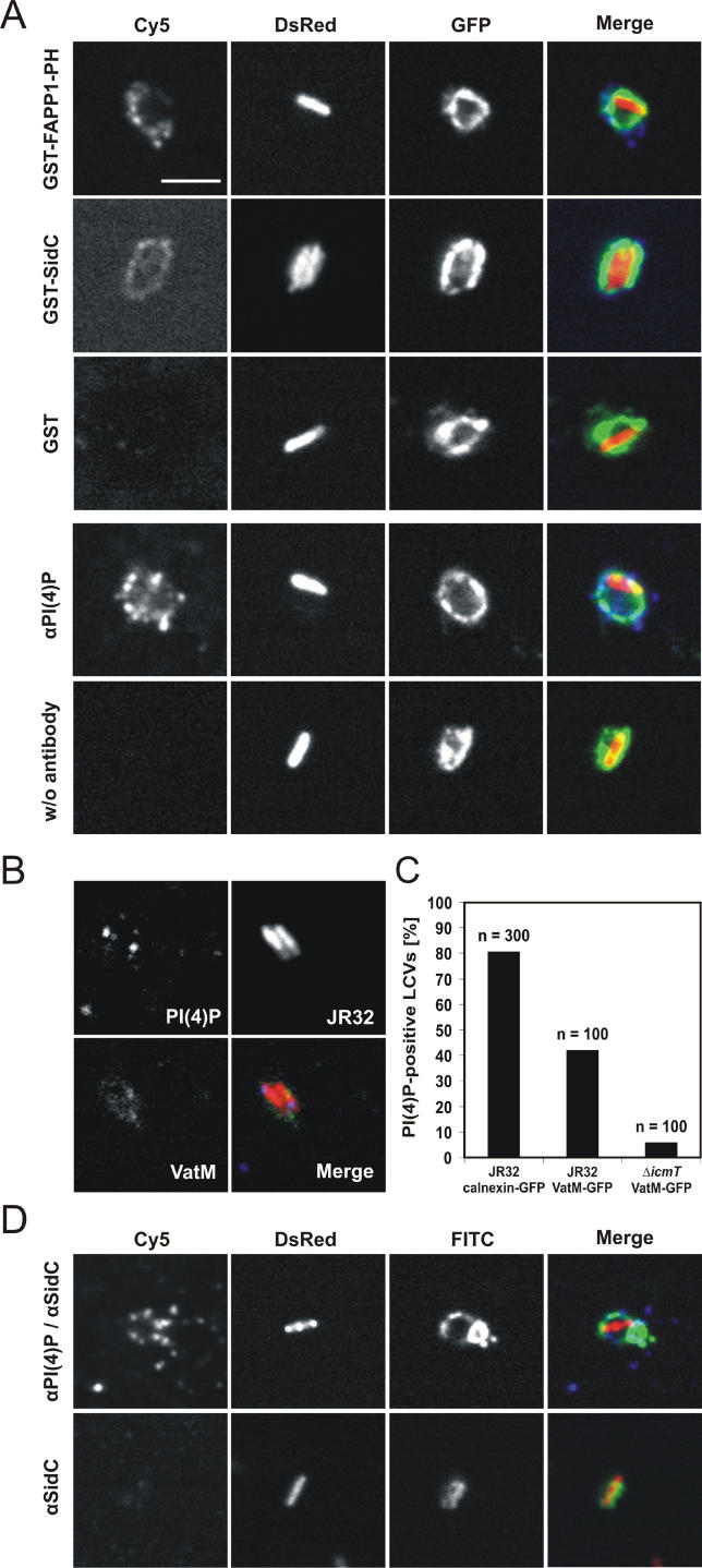
The causative agent of Legionnaires' disease, Legionella pneumophila, employs the intracellular multiplication (Icm)/defective organelle trafficking (Dot) type IV secretion system (T4SS) to upregulate phagocytosis and to establish a replicative vacuole in amoebae and macrophages. Legionella-containing vacuoles (LCVs) do not fuse with endosomes but recruit early secretory vesicles. Here we analyze the role of host cell phosphoinositide (PI) metabolism during uptake and intracellular replication of L. pneumophila. Genetic and pharmacological evidence suggests that class I phosphatidylinositol(3) kinases (PI3Ks) are dispensable for phagocytosis of wild-type L. pneumophila but inhibit intracellular replication of the bacteria and participate in the modulation of the LCV. Uptake and degradation of an icmT mutant strain lacking a functional Icm/Dot transporter was promoted by PI3Ks. We identified Icm/Dot-secreted proteins which specifically bind to phosphatidylinositol(4) phosphate (PI(4)P) in vitro and preferentially localize to LCVs in the absence of functional PI3Ks. PI(4)P was found to be present on LCVs using as a probe either an antibody against PI(4)P or the PH domain of the PI(4)P-binding protein FAPP1 (phosphatidylinositol(4) phosphate adaptor protein-1). Moreover, the presence of PI(4)P on LCVs required a functional Icm/Dot T4SS. Our results indicate that L. pneumophila modulates host cell PI metabolism and exploits the Golgi lipid second messenger PI(4)P to anchor secreted effector proteins to the LCV.


Conflict of interest statement

Competing interests. A provisional US patent application has been granted to Hubert Hilbi (US60/719,934: “Bacterial phosphoinositide probes and effectors”).

Figures

Figure 1
Figure 1. Phagocytosis of Wild-Type L. pneumophila or ΔicmT by Dictyostelium
Phagocytosis of gfp-expressing wild-type L. pneumophila (black bars) or ΔicmT (grey bars) by Dictyostelium infected at (A and B) an MOI of 100 or (C) at the MOI indicated was analyzed by flow cytometry. The increase in GFP fluorescence (FL1, x-axis) indicates that, at MOIs ranging from 1–100, the number of wild-type L. pneumophila phagocytosed is about one order of magnitude higher than the number of ΔicmT. Phagocytosis is blocked by latrunculin B or incubation at 4 °C. (B and C) The data shown are the means and standard deviations of duplicates and are representative of at least three independent experiments.
Figure 2
Figure 2. A Role for PI3Ks in Intracellular Replication but Not Phagocytosis of Wild-Type L. pneumophila
(A) Phagocytosis by Dictyostelium wild-type Ax3 or ΔPI3K1/2 (untreated or treated with 5 μM WM) of GFP-labeled L. pneumophila wild-type (black bars) or a ΔicmT mutant strain (grey bars) was determined by flow cytometry. (B) Release of L. pneumophila wild-type (squares) or ΔicmT (circles) into the supernatant of Dictyostelium wild-type (denoted by filled symbols) or ΔPI3K1/2 (denoted by open symbols) was quantified by CFUs. (C) Release of wild-type L. pneumophila (CFUs) from Dictyostelium wild-type (filled squares denote untreated; filled triangles denote 10 μM LY) or ΔPI3K1/2 (open squares denote untreated; open triangles denote LY). (D) Quantification by flow cytometry of intracellular growth of GFP-labeled wild-type L. pneumophila within wild-type Dictyostelium or ΔPI3K1/2 in the presence or absence of 20 μM LY. The data shown are means and standard deviations of duplicates (A) or triplicates (B and C), and are representative of at least three independent experiments (A–D).
Figure 3
Figure 3. A Role for PI3Ks in Degradation of L. pneumophila ΔicmT
Quantification by flow cytometry of intracellular degradation of GFP-labeled L. pneumophila ΔicmT (MOI 100, MOI 500) within wild-type Dictyostelium or ΔPI3K1/2. In the absence of PI3Ks, ΔicmT is less efficiently phagocytosed and degraded.
Figure 4
Figure 4. Trafficking of L. pneumophila within Dictyostelium Lacking Functional PI3Ks
(A) Confocal laser scanning micrographs of calnexin-GFP–labeled Dictyostelium (green) wild-type Ax3 (WT denotes untreated or treated with 20 μM LY) or ΔPI3K1/2 (denoted by ΔPI3K) infected with DsRed-Express–labeled wild-type L. pneumophila (red) for 1 h or 2.5 h, respectively. Representative examples of spacious vacuoles (WT) and tight vacuoles (WT/LY, ΔPI3K) are shown. DNA was stained with DAPI (blue). Bar denotes 2 μm. (B) Quantification over time of spacious LCVs in calnexin-GFP–labeled Dictyostelium wild-type (filled squares denote untreated; filled triangles denote LY) or ΔPI3K1/2 (open squares) infected with DsRed-Express–labeled wild-type L. pneumophila. Data represent means and standard deviations of the percentage of spacious vacuoles from 50–200 total vacuoles per time point scored in four independent experiments. (C) Confocal laser scanning micrographs of Dictyostelium wild-type or ΔPI3K1/2, infected for 75 min with wild-type L. pneumophila expressing M45-tagged SidC. Infected amoebae were stained with rhodamine-conjugated anti-L. pneumophila antibody (red), FITC-conjugated anti-M45-tag antibody (green), and DAPI (blue), respectively. Bar denotes 2 μm.
Figure 5
Figure 5. Binding of L. pneumophila Icm/Dot–Secreted Proteins to PIs In Vitro
(A) Binding of affinity-purified GST fusion proteins of SidC, SdcA, SidD, or SdhB (160 pmol) to different lipids (100 pmol; left panels) or 2-fold serial dilutions of PIs (100–1.56 pmol; right panels) immobilized on nitrocellulose membranes was analyzed by a protein-lipid overlay assay using an anti-GST antibody. Lysophosphatidic acid is denoted by LPA; lysophosphocholine is denoted by LPC; sphingosine-1-phosphate is denoted by SP; phosphatidic acid is denoted by PA; and phosphatidylserine is denoted by PS. The experiment was reproduced at least three times with similar results. (B) PL vesicles (20 μl, 1 mM lipid) composed of PC (65%), PE (30%), and 5% (1 nmol) either PI(4)P, PI(3)P or PI(4,5)P2 were incubated with affinity-purified GST-SidC or GST-SidD (40 pmol), centrifuged, and washed. Binding of GST fusion proteins to PL vesicles was assayed by Western blot with an anti-GST antibody. Similar results were obtained in three separate experiments.
Figure 6
Figure 6. PI3Ks Affect the Amount of SidC Bound to LCVs in Dictyostelium
(A) Confocal laser scanning micrographs of calnexin-GFP–labeled Dictyostelium wild-type strain Ax3 (green), infected with DsRed-Express–labeled wild-type L. pneumophila (red) for 1 h (left panel), and immuno-labeled for SidC (blue) with an affinity-purified primary and Cy5-conjugated secondary antibody (middle panel). To quantify fluorescence intensity (right panel), the averaged fluorescence intensity of background areas (B1, B2, and B3) was subtracted from the intensity of the sample area (S). Bar denotes 2 μm. (B) Dot plot of SidC fluorescence (average and variance) on LCVs within Dictyostelium wild-type (untreated, n = 135; 20 μM LY, n = 94) or ΔPI3K1/2 (n = 86). The data shown are combined from six independent experiments, each normalized to 100% (average SidC fluorescence on LCVs in wild-type Dictyostelium).
Figure 7
Figure 7. PI(4)P Is a Lipid Marker of LCVs Harboring Icm/Dot–Proficient L. pneumophila
(A, B, and D) Confocal micrographs of LCVs in lysates of (A) calnexin-GFP–labeled Dictyostelium, (B) VatM-GFP–labeled Dictyostelium, or (D) RAW264.7 macrophages infected with DsRed-Express–labeled L. pneumophila are shown. The lysates were prepared with a ball homogenizer, and PI(4)P was visualized on the LCVs using as probes either the PH domain of the PI(4)P-binding protein FAPP1 fused to GST, an antibody against PI(4)P, or GST-SidC. Using GST alone or omission of the anti-PI(4)P antibody did not label the LCVs. Bar denotes 2 μm (magnification of all images is identical). (C) Quantification of PI(4)P-positive calnexin-GFP–labeled (n = 300) or VatM-GFP–labeled (n = 100) LCVs in Dictyostelium wild-type strain Ax3.

References

    1. Fields BS. The molecular ecology of Legionellae . Trends Microbiol. 1996;4:286–290. - PubMed
    1. McDade JE, Shepard CC, Fraser DW, Tsai TR, Redus MA, et al. Legionnaires' disease: Isolation of a bacterium and demonstration of its role in other respiratory disease. N Engl J Med. 1977;297:1197–1203. - PubMed
    1. Nash TW, Libby DM, Horwitz MA. Interaction between the Legionnaires' disease bacterium (Legionella pneumophila) and human alveolar macrophages. Influence of antibody, lymphokines, and hydrocortisone. J Clin Invest. 1984;74:771–782. - PMC - PubMed
    1. Horwitz MA. The Legionnaires' disease bacterium (Legionella pneumophila) inhibits lysosome-phagosome fusion in human monocytes. J Exp Med. 1983;158:2108–2126. - PMC - PubMed
    1. Kagan JC, Roy CR. Legionella phagosomes intercept vesicular traffic from endoplasmic reticulum exit sites. Nat Cell Biol. 2002;4:945–954. - PubMed

Publication types

MeSH terms

Substances