. 2006 Jun;116(6):1615-23.
doi: 10.1172/JCI27238. Epub 2006 May 18.

Neutrophils and their Fc gamma receptors are essential in a mouse model of transfusion-related acute lung injury

Affiliations

Neutrophils and their Fc gamma receptors are essential in a mouse model of transfusion-related acute lung injury

Mark R Looney et al. J Clin Invest. 2006 Jun.

Abstract

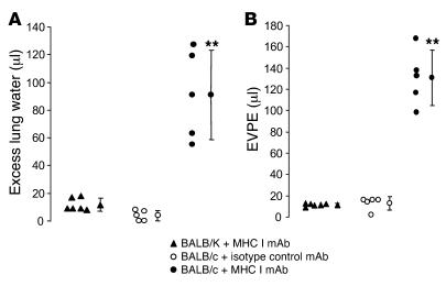
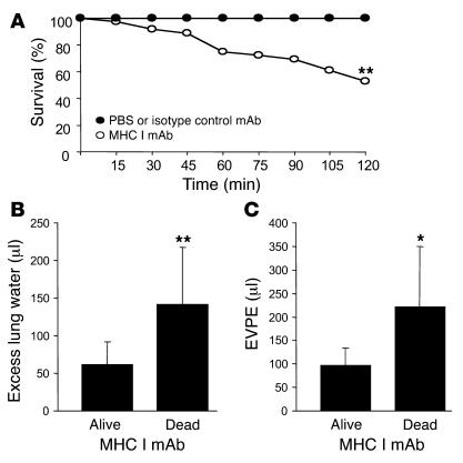
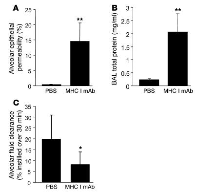
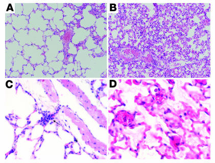
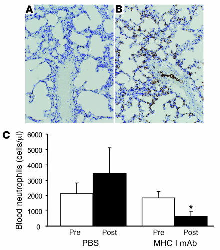
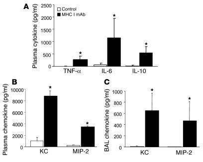
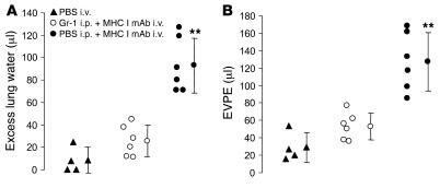
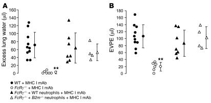
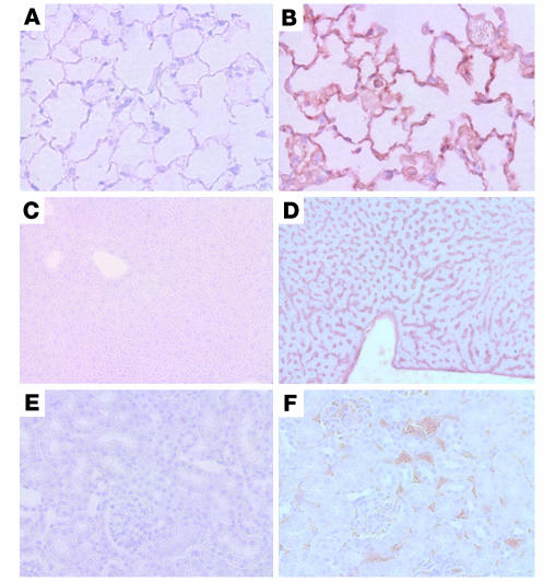
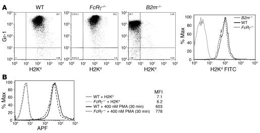
Transfusion-related acute lung injury (TRALI) is the most common cause of transfusion-related mortality. To explore the pathogenesis of TRALI, we developed an in vivo mouse model based on the passive transfusion of an MHC class I (MHC I) mAb (H2Kd) to mice with the cognate antigen. Transfusion of the MHC I mAb to BALB/c mice produced acute lung injury with increased excess lung water, increased lung vascular and lung epithelial permeability to protein, and decreased alveolar fluid clearance. There was 50% mortality at a 2-hour time point after Ab administration. Pulmonary histology and immunohistochemistry revealed prominent neutrophil sequestration in the lung microvasculature that occurred concomitantly with acute peripheral blood neutropenia, all within 2 hours of administration of the mAb. Depletion of neutrophils by injection of anti-granulocyte mAb Gr-1 protected mice from lung injury following MHC I mAb challenge. FcRgamma-/- mice were resistant to MHC I mAb-induced lung injury, while adoptive transfer of wild-type neutrophils into the FcRgamma-/- animals restored lung injury following MHC I mAb challenge. In conclusion, in a clinically relevant in vivo mouse model of TRALI using an MHC I mAb, the mechanism of lung injury was dependent on neutrophils and their Fc gamma receptors.


Figures

Figure 1
Figure 1. MHC I mAb produces ALI in BALB/c mice.
Gravimetric excess lung water (A) and EVPE determined from i.v. 125I-labeled albumin protein leakage (B) were increased in BALB/c mice (H2Kd) given MHC I mAb (4.5 mg/kg) but not in BALB/K controls or in BALB/c mice given an isotype-matched Ab (IgG2a, κ). Each data point represents a single animal. **P < 0.01 compared with BALB/K + MHC I mAb and BALB/c + isotype control mAb.
Figure 2
Figure 2. MHC I mAb produces mortality and increased lung injury in nonsurvivors versus survivors.
(A) BALB/c mice given MHC I mAb (n = 36) showed decreased survival at 2 hours compared with BALB/c mice given either isotype-matched mAb or PBS (n = 22). **P < 0.01, c2 test. The MHC I mAb–challenged mice that died before 2 hours (n = 15) had increased excess lung water (B) and increased EVPE (C) compared with MHC I mAb–challenged mice that survived the 2-hour experimental period (n = 15). **P < 0.01; *P < 0.05.
Figure 3
Figure 3. MHC I mAb produces increased alveolar epithelial permeability and decreased alveolar fluid clearance.
(A and B) Alveolar epithelial permeability to 125I-labeled albumin (A) and BAL total protein (B) in BALB/c mice given PBS (n = 6) or MHC I mAb (n = 10). **P < 0.01. (C) In situ alveolar fluid clearance was measured over 30 minutes in mice treated with MHC I mAb (n = 7) or PBS (n = 6). *P < 0.05.
Figure 4
Figure 4. Lung histology from control and MHC I mAb–treated mice.
(A and B) Low-power views of lungs from mice given either control (A) or MHC I mAb (B). In the MHC I mAb–treated mouse, there was increased intravascular neutrophils, septal thickening, and interstitial inflammation. (C) High-power view of lung from a mouse given MHC I mAb. Arrow indicates a branching vessel that is plugged with neutrophils. (D) MHC I mAb–treated mouse with intra-alveolar proteinaceous material. H&E staining. Magnification, ×200 (A and B); ×400 (C and D).
Figure 5
Figure 5. MHC I mAb–treated mice develop lung neutrophil sequestration and neutropenia.
(A and B) Low-power view of lung from BALB/c mouse given either PBS (A) or MHC I mAb (B) and stained with Gr-1 anti-neutrophil mAb. Note the extensive neutrophil sequestration (brown color) in the MHC I mAb–treated mouse lung. Magnification, ×200. (C) Neutrophil levels in the blood. Two hours after injection of either PBS or MHC I mAb compared with baseline preinjection levels. *P < 0.05.
Figure 6
Figure 6. MHC I mAb–induced lung injury is characterized by an acute inflammatory and antiinflammatory response and an increase in plasma and BAL chemokines.
(A) Plasma samples obtained 2 hours after anti-MHC I mAb treatment showed higher levels of inflammatory cytokines TNF-α and IL-6 as well as elevated levels of IL-10 compared with controls (n = 5 per group). (B and C) KC and MIP-2 levels were also increased in the plasma (2 hours after MHC I mAb treatment) (B) and the BAL (C) of MHC I mAb–treated mice (n = 6 per group). *P < 0.01.
Figure 7
Figure 7. Neutrophil depletion with Gr-1 mAb protects mice from MHC I mAb–induced ALI.
Mice were pretreated with either i.p. PBS or i.p. Gr-1 mAb (250 μg) and after 24 hours were given i.v. MHC I mAb and compared with mice given i.v. PBS. The Gr-1 mAb–pretreated mice had greatly attenuated excess lung water (A) and lung vascular permeability to 125I-labeled albumin (B) compared with mice pretreated with PBS. Each data point represents an individual animal. **P < 0.01.
Figure 8
Figure 8. FcRγ–/– mice were protected from MHC I mAb–induced ALI and adoptive transfer of wild-type, andB2m–/– neutrophils restored lung injury inFcRγ–/– mice.
FcRγ–/–mice, wild-type matched controls (BALB/c), and FcRγ–/–mice injected with 5 × 10⁶ wild-type or B2m–/–neutrophils were given i.v. MHC I mAb, and excess lung water (A) and lung vascular permeability (B) were measured. Each data point represents an individual animal. **P < 0.01, FcRγ–/–versus wild-type and FcRγ–/–plus wild-type or B2m–/–neutrophils.
Figure 9
Figure 9. Distribution of MHC I mAb after i.v. injection.
Rag2–/– mice were injected i.v. with PBS (A, C, and E) or 4.5 mg/kg MHC I mAb (B, D, and F) and sacrificed after 2 hours. The lungs (A and B; magnification, ×600), liver (C and D; magnification, ×200), and kidney (E and F; magnification, ×400) were examined.
Figure 10
Figure 10. MHC I mAb binds but does not activate wild-type andFcRγ–/– neutrophils.
(A) Flow cytometric analysis of MHC I expression on bone marrow neutrophils. Neutrophils were obtained from the bone marrow and stained with anti-H2Kd and the granulocyte-specific marker Gr-1. In B2m–/– mice, the cells were negative for H2Kd staining. (B) Flow cytometric analysis of superoxide production by bone marrow neutrophils. Superoxide anion levels were determined with an aminophenyl fluorescein (APF) flow cytometry assay. Mouse bone marrow neutrophils were either stimulated by MHC I cross-linking (25 mg/ml anti–H2Kd-biotin followed by 40 mg/ml streptavidin) or 400 nM PMA. Representative results from individual mice are shown.

Similar articles

Cited by

References

    1. Toy P., et al. Transfusion-related acute lung injury: definition and review. Crit. Care Med. 2005;33:721–726. - PubMed
    1. Looney M.R., Gropper M.A., Matthay M.A. Transfusion-related acute lung injury: a review. Chest. 2004;126:249–258. - PubMed
    1. Kopko P.M., Marshall C.S., MacKenzie M.R., Holland P.V., Popovsky M.A. Transfusion-related acute lung injury: report of a clinical look-back investigation. JAMA. 2002;287:1968–1971. - PubMed
    1. Kleinman S., et al. Toward an understanding of transfusion-related acute lung injury: statement of a consensus panel. Transfusion. 2004;44:1774–1789. - PubMed
    1. Popovsky M.A., Moore S.B. Diagnostic and pathogenetic considerations in transfusion-related acute lung injury. Transfusion. 1985;25:573–577. - PubMed

Publication types

MeSH terms