Review
. 2006 Jun;16(3):279-87.
doi: 10.1016/j.sbi.2006.05.009. Epub 2006 May 19.

The building blocks and motifs of RNA architecture

Affiliations
Review

The building blocks and motifs of RNA architecture

Neocles B Leontis et al. Curr Opin Struct Biol. 2006 Jun.

Abstract

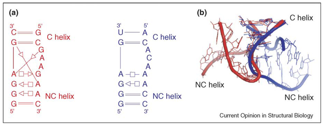
RNA motifs can be defined broadly as recurrent structural elements containing multiple intramolecular RNA-RNA interactions, as observed in atomic-resolution RNA structures. They constitute the modular building blocks of RNA architecture, which is organized hierarchically. Recent work has focused on analyzing RNA backbone conformations to identify, define and search for new instances of recurrent motifs in X-ray structures. One current view asserts that recurrent RNA strand segments with characteristic backbone configurations qualify as independent motifs. Other considerations indicate that, to characterize modular motifs, one must take into account the larger structural context of such strand segments. This follows the biologically relevant motivation, which is to identify RNA structural characteristics that are subject to sequence constraints and that thus relate RNA architectures to sequences.


Figures

Figure 1
Figure 1
Five examples of the new Ω-turn motif, as proposed by Wadley and Pyle [47••], in their structural contexts: (a) 23S rRNA from H. marismortui (PDB code 1JJ2), (b) 16S rRNA from Thermus thermophilus (PDB code 1N32), (c) RNA aptamer (PDB code 1NTB), (d) 23S rRNA from H. marismortui (PDB code 1JJ2) and (e) 23S rRNA from H. marismortui (PDB code 1JJ2). The upper panel shows 3D representations highlighting the conservation of the backbone conformation of the five nucleotides composing each Ω-turn (shown in red). The lower panel shows schematic representations of each Ω-turn in its structural context, annotated with symbols for base-pairing [31] and base-stacking [48] interactions. The nucleotides composing each Ω-turn are shown in red and nucleotides in the syn conformation are indicated in bold. To prepare Figure 1, each Ω-turn was visually inspected in its structural context, and annotated for base-pairing [31] and base-stacking [48] interactions. This analysis clearly shows that motifs comprising Ω-turns share other common structural characteristics and are subject to sequence constraints. Thus, the first base of each Ω-turn forms a WC base pair. In three out of the five cases reported, the second base forms a trans Hoogsteen/sugar edge (sheared) pair [47••]. In the third case, the second base is a uridine, which cannot make a trans Hoogsteen/sugar edge pair, but rather interacts with C18 to form a cis Hoogsteen/sugar edge pair. However, the position of this second base is the same as in the other Ω-turns. In the fifth case, the corresponding base (G1417 in H. marismortui 23S rRNA) is in the syn glycosidic configuration and, were it to rotate back to the more common anti configuration, it would form the same type of base pair with A1678. In each Ω-turn, the fourth base is in the syn glycosidic configuration and is extruded from the helix formed by the preceding nucleotides, so as to form a cis WC base pair with the base belonging to the other strand that pairs with the second base of the Ω-turn strand. The third base also base pairs, in a variable fashion, but always forming a trans base pair.
Figure 2
Figure 2
Kink-turn and reverse kink-turn. (a) Schematic structures of the helix 7 kink-turn in 23S rRNA from H. marismortui (shown in red) and the reverse kink-turn in the P9/P9.0 junction of the Azoarcus intron (shown in blue). The structures are annotated for base-pairing interactions using the geometric nomenclature of Leontis and Westhof [31]. (b) Superposition of the canonical helices (C helix) from the 3D structures of the kink-turn (red) and the reverse kink-turn (blue). In the kink-turn structure, the non-canonical helix (NC helix) is oriented in the minor/shallow groove, whereas in the reverse kink-turn structure, the NC helix is in the major/deep groove.
Figure 3
Figure 3
Flow chart illustrating the use of isostericity matrices to integrate 3D structural and sequence information to produce accurate alignments and model 3D structures based on sequence. Isostericity matrices for non-WC base pairs organized in geometric families were proposed based on analysis of high-resolution atomic structures [54], as indicated in path 1. Sequence signatures of RNA motifs identified in 3D structures are deduced by analyzing homologous RNA molecules that have the same motif (path 2). Isostericity matrices are employed to productively iterate between sequence alignment and sequence signature to arrive at accurate, structure-based alignments (path 2). Sequence signatures for recurrent motifs identified in different crystal structures are defined with reference to isostericity matrices (path 3). For families of homologous RNA molecules for which no 3D structure exists (path 4), WC covariations and energy minimization (path 5) can be used to determine common 2D structures, which in turn define hairpin, internal and junction loops in which 3D motifs may occur. Sequence signatures of known motifs are used to propose motifs for loops and to refine alignments of loop regions in an iterative manner (paths 4 and 5). Motif substitutions at corresponding positions in the alignments can also be identified (path 4).

References

    1. Leontis NB, Westhof E. Analysis of RNA motifs. Curr Opin Struct Biol. 2003;13:300–308. - PubMed
    1. Holbrook SR. RNA structure: the long and the short of it. Curr Opin Struct Biol. 2005;15:302–308. Recently determined crystallographic structures are reviewed. All new and recurrent RNA motifs in recent structures are identified and described.

    1. Khusial P, Plaag R, Zieve GW. LSm proteins form heptameric rings that bind to RNA via repeating motifs. Trends Biochem Sci. 2005;30:522–528. - PubMed
    1. Mathews DM, Zuker M. Predictive methods using RNA sequences. In: Baxevanis AD, Ouellette BFF, editors. Bioinformatics: A Practical Guide to the Analysis of Genes and Proteins. John Wiley & Sons; 2005. pp. 144–171.
    1. Zorn J, Gan HH, Shiffeldrim N, Schlick T. Structural motifs in ribosomal RNAs: implications for RNA design and genomics. Biopolymers. 2004;73:340–347. - PubMed

Publication types

LinkOut - more resources