. 2006 May 30;103(22):8459-64.
doi: 10.1073/pnas.0603082103. Epub 2006 May 19.

Essential role of mda-5 in type I IFN responses to polyriboinosinic:polyribocytidylic acid and encephalomyocarditis picornavirus

Affiliations

Essential role of mda-5 in type I IFN responses to polyriboinosinic:polyribocytidylic acid and encephalomyocarditis picornavirus

Leonid Gitlin et al. Proc Natl Acad Sci U S A. .

Abstract

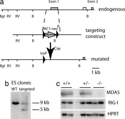
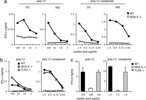
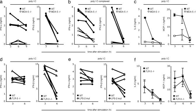
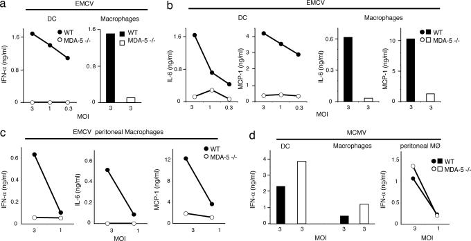
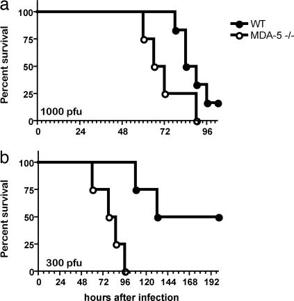
The innate immune system recognizes viral dsRNA through two distinct pathways; the Toll-like receptor 3 (TLR3) pathway detects dsRNA phagocytosed in endosomes; the helicases retinoic acid-induced protein I (RIG-I) and melanoma differentiation-associated gene-5 (mda-5) detect cytoplasmic dsRNA generated during viral replication. Both RIG-I and mda-5 can bind polyriboinosinic:polyribocytidylic acid (polyI:C), the synthetic analog of viral dsRNA, and mediate type I IFN responses to polyI:C and multiple RNA viruses in vitro. We generated mda-5-deficient mice and showed that mda-5 is the dominant receptor mediating type I IFN secretion in response to polyI:C in vitro and in vivo. Moreover, mda-5-/- mice exhibited a selectively impaired antiviral response to encephalomyocarditis picornavirus, indicating functional specialization of mda-5 in vivo.


Conflict of interest statement

Conflict of interest statement: No conflicts declared.

Figures

Fig. 1.
Fig. 1.
Generation of mda-5−/−mice. (a) Map of the endogenous mda-5 allele, targeting construct, and the mutated mda-5 allele after the removal of MC1-neoR by Cre recombination. mda-5 exons are shown as gray boxes, the external probe as an open box, and the MC1-neoR construct as an arrow. LoxP sites are designated by filled arrowheads. Restriction enzyme sites: B, BamHI; Bgl, BglII; and RV, EcoRV. (b) DNA blot analysis of ES cell DNA. Genomic DNA was cut with BamHI, and the blot was hybridized with the probe indicated in a. (c) RNA blot analysis of total liver and kidney RNA of WT, heterozygous, and mda-5−/− littermates. Mice were injected with polyI:C 3 days before collection of organs. For each mouse, the left lane represents liver, and the right lane represents kidney. mda-5 (Top), RIG-I (Middle), and hypoxanthine-guanine phosphoribosyl transferase (HPRT) (Bottom) blots are shown.
Fig. 2.
Fig. 2.
mda-5 is critically required for the type I IFN response of bone marrow-derived DC and macrophages to polyI:C. DC and macrophages derived from WT (filled symbols), mda-5-deficient (open symbols, a–c), and TLR3-deficient mice (gray symbols, b and c) were stimulated in vitro, as indicated, with synthetic polyI:C or with polyI:C in complex with Fugene. Cell culture supernatants were assessed after 24 h of stimulation by ELISA for IFN-α (a and b) and IFN-β (c). Data shown are representative of four (a) and two (b and c) independent experiments.
Fig. 3.
Fig. 3.
Distinct and complementary contributions of mda-5 and TLR3-TRIF to polyI:C-induced responses in thioglycollate-induced peritoneal macrophages. Macrophages (2 × 10⁵ per well) from WT and TRIFLPS2 mice (a) or WT and mda-5−/− mice (b) were collected 48 h after i.p. thioglycollate injection and stimulated with polyI:C either naked (100 μg/ml) or in complex with Fugene (1.5 μg/ml). After 24 h, cell culture supernatants were assessed for IFN-α by ELISA and for TNF-α, MCP-1, and IL-6 by cytokine bead array. Data shown are representative of two independent experiments.
Fig. 4.
Fig. 4.
mda-5 deficiency strongly impairs the type I IFN response to polyI:C in vivo. WT, mda-5-deficient, TLR3-deficient, or TRIFLPS-2 (LPS-2 mutant) mice were injected i.v., as indicated, with either 100 μg of polyI:C or 10 μg of polyI:C complexed with Fugene. Serum samples were taken 3, 6, and 9 h after stimulation and were analyzed by ELISA for IFN-α and IFN-β (a, b, d, and e). Lines show cytokine kinetics in individual mice. IL-6 and MCP-1 were assessed by cytokine bead array (c and f). Error bars indicate SEM.
Fig. 5.
Fig. 5.
mda-5 is critically required for type I IFN and cytokine response to EMCV. Bone marrow-derived DC (a, b, and d), bone marrow-derived macrophages (a, b, and d), and thioglycollate-induced peritoneal macrophages (c and d) from WT (filled symbols) and mda-5-deficient (open symbols) mice were stimulated in vitro with EMCV or MCMV at indicated multiplicities of infection. Production of IFN-α (a and c) and IL-6 and MCP-1 (b and c) in response to EMCV. (d) IFN-α response to MCMV. Cell culture supernatants were examined for IFN-α by ELISA and for IL-6 and MCP-1 by cytokine bead array. Data shown are representative of two independent experiments.
Fig. 6.
Fig. 6.
mda-5-deficient mice show increased susceptibility to lethal infections with EMCV. (a) mda-5−/− mice (n = 4, open symbols) and WT littermate controls (n = 6, filled symbols) on a pure 129 SvJ background were injected i.v. with 1,000 plaque-forming units (pfu) of EMCV and then monitored for survival. mda-5−/− mice died after a mean survival time of 69 h, whereas WT mice survived for a mean of 87 h (P = 0.07). (b) mda-5−/− mice (n = 5, open symbols) and WT littermate controls (n = 5, filled symbols) on a B6x129 SvJ background were injected i.v. with 300 pfu of EMCV and monitored for survival. mda-5−/− mice died after a mean survival time of 82 h, whereas WT mice survived for a mean of 166 h (P = 0.008).

References

    1. Janeway C. A., Jr, Medzhitov R. Annu. Rev. Immunol. 2002;20:197–216. - PubMed
    1. Akira S., Uematsu S., Takeuchi O. Cell. 2006;124:783–801. - PubMed
    1. Alexopoulou L., Holt A. C., Medzhitov R., Flavell R. A. Nature. 2001;413:732–738. - PubMed
    1. Matsumoto M., Funami K., Tanabe M., Oshiumi H., Shingai M., Seto Y., Yamamoto A., Seya T. J. Immunol. 2003;171:3154–3162. - PubMed
    1. Schulz O., Diebold S. S., Chen M., Naslund T. I., Nolte M. A., Alexopoulou L., Azuma Y. T., Flavell R. A., Liljestrom P., Reis e Sousa C. Nature. 2005;433:887–892. - PubMed

Publication types

MeSH terms

LinkOut - more resources