. 2006 May 19;34(9):2723-35.
doi: 10.1093/nar/gkl348. Print 2006.

Human telomeric sequence forms a hybrid-type intramolecular G-quadruplex structure with mixed parallel/antiparallel strands in potassium solution

Affiliations

Human telomeric sequence forms a hybrid-type intramolecular G-quadruplex structure with mixed parallel/antiparallel strands in potassium solution

Attila Ambrus et al. Nucleic Acids Res. .

Abstract

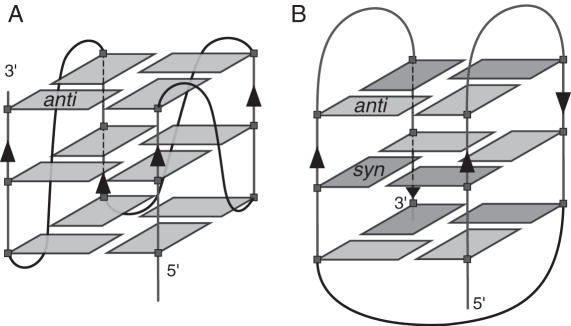
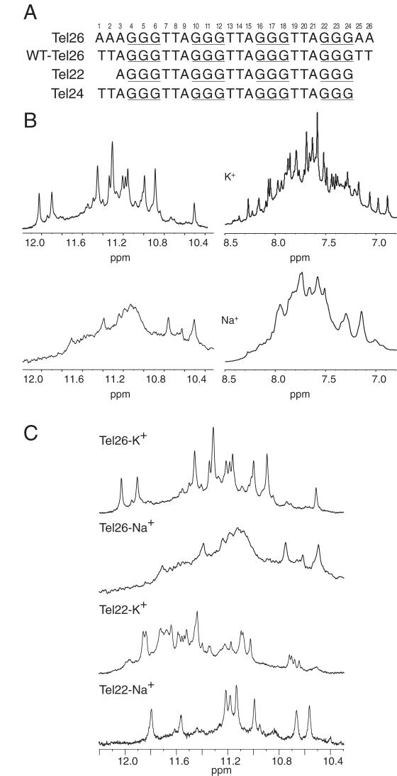
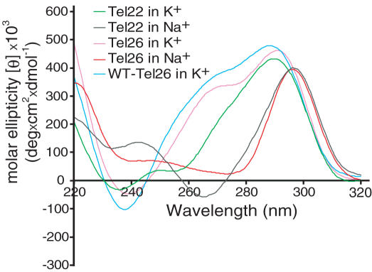
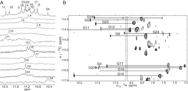
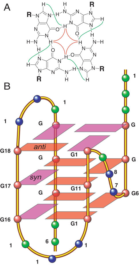
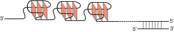
Human telomeric DNA consists of tandem repeats of the sequence d(TTAGGG). The formation and stabilization of DNA G-quadruplexes in the human telomeric sequence have been shown to inhibit the activity of telomerase, thus the telomeric DNA G-quadruplex has been considered as an attractive target for cancer therapeutic intervention. However, knowledge of the intact human telomeric G-quadruplex structure(s) formed under physiological conditions is a prerequisite for structure-based rational drug design. Here we report the folding structure of the human telomeric sequence in K+ solution determined by NMR. Our results demonstrate a novel, unprecedented intramolecular G-quadruplex folding topology with hybrid-type mixed parallel/antiparallel G-strands. This telomeric G-quadruplex structure contains three G-tetrads with mixed G-arrangements, which are connected consecutively with a double-chain-reversal side loop and two lateral loops, each consisting of three nucleotides TTA. This intramolecular hybrid-type telomeric G-quadruplex structure formed in K+ solution is distinct from those reported on the 22 nt Tel22 in Na+ solution and in crystalline state in the presence of K+, and appears to be the predominant conformation for the extended 26 nt telomeric sequence Tel26 in the presence of K+, regardless of the presence or absence of Na+. Furthermore, the addition of K+ readily converts the Na+-form conformation to the K+-form hybrid-type G-quadruplex. Our results explain all the reported experimental data on the human telomeric G-quadruplexes formed in the presence of K+, and provide important insights for understanding the polymorphism and interconversion of various G-quadruplex structures formed within the human telomeric sequence, as well as the effects of sequence and cations. This hybrid-type G-quadruplex topology suggests a straightforward pathway for the secondary structure formation with effective packing within the extended human telomeric DNA. The hybrid-type telomeric G-quadruplex is most likely to be of pharmacological relevance, and the distinct folding topology of this G-quadruplex suggests that it can be specifically targeted by G-quadruplex interactive small molecule drugs.


Figures

Figure 1
Figure 1
Folding topologies of Tel22. (A) Propeller-type parallel-stranded intramolecular G-quadruplex in the presence of K+ in crystalline state. (B) Basket-type mixed parallel/antiparallel-stranded intramolecular G-quadruplex in Na+ solution determined by NMR.
Figure 2
Figure 2
(A) Different four repeat telomeric DNA sequences. WT-Tel26, Tel22 and Tel24 are the wild-type four repeat telomeric DNA sequences with sizes of 26, 22 and 24 nt, respectively. The numbering system is shown above Tel26. (B) Imino and aromatic regions of 1D 1H NMR spectra of Tel26 in K+ and Na+ solutions at 25°C. (C) Imino regions of 1D 1H NMR spectra of Tel22 and Tel26 in Na+ and K+ solutions at 25°C.
Figure 3
Figure 3
CD spectra of Tel26, WT-Tel26 and Tel22 in 100 mM Na+ or K+ solutions at 25°C. The same CD signatures are observed for Tel26 and WT-Tel26 in K+ solution, while similarly distinct CD signatures are observed for Tel22 and Tel26 in Na+ solution.
Figure 4
Figure 4
(A) Imino proton assignments of Tel26 using 1D 15N-filtered experiments on site-specific labeled oligonucleotides. Conditions: 25 mM K-PO4, 70 mM KCl, pH 7.0, 25°C, 0.6–0.7 mM DNA. (B) Long-range through-bond correlations between intraresidue imino and H8 protons via C5 of the guanine base, determined by JR-HMBC experiment at natural abundance (46).
Figure 5
Figure 5
(A) A G-tetrad with H1–H1 and H1–H8 connectivity patterns detectable in NOESY experiments. (B) Schematic diagram of the folding topology of the unimolecular human telomeric G-quadruplex in K+ solution. red ball, guanine; red box, (anti) guanine; magenta box, (syn) guanine; green ball, adenine; blue ball, thymine.
Figure 6
Figure 6
The expanded H1–H8/H2/H6 and H1–H1 regions of the exchangeable proton 2D JR-NOESY spectrum of Tel26 in K+ solution at 1°C. NOEs are labeled as follows: intra-tetrad NOEs are labeled in red, sequential NOEs are labeled in green, inter-tetrad NOEs are labeled in blue, NOEs between stacking adenine H2 protons and G-tetrad imino protons are labeled in black. Conditions: 1°C, 25 mM K-PO4, 70 mM KCl, pH 7.0, 2.5 mM DNA.
Figure 7
Figure 7
Titration experiments of K+ in the presence of 150 mM Na+ for Tel26 (A) and Tel22 (C), and titration experiments of Na+ in the presence of 100 mM K+ for Tel26 (B) and Tel22 (D), monitored by CD spectroscopy.
Figure 8
Figure 8
(A) Imino proton region of Tel26 in K+ solution in a variable temperature study. As seen above 65°C there is no detectable peak intensity in the imino (exchangeable proton) region. The imino protons are labeled on the spectrum recorded at 20°C. (B) D2O-to-H2O NMR exchange experiments. The imino region of the 1D 1H NMR spectrum of Tel26, which was first incubated overnight in 99.96% D2O and then incubated in H2O for 30 min, 3.5 h and then overnight at 25°C. Imino protons from the middle layer of the G-tetrad (Figure 5) are the last to appear and are labeled with the residue numbers. (C) Melting profile of Tel26 in K+ solution in a CD variable temperature study. The same melting temperature of 55°C is shown by both CD and NMR (A).
Figure 9
Figure 9
Comparison of G-quadruplex-forming sequences. Loops colored in red have been shown to adopt parallel-stranded double-chain-reversal side loops (for more details see text).
Figure 10
Figure 10
Schematic diagram of interconversions between the Na+ and K+ forms of telomeric G-quadruplexes. (A) For the extended four repeat telomeric sequence Tel26, the hybrid-type G-quadruplex structure is the most stable and thus the predominant form in the presence of K+, regardless of the presence or absence of Na+. Addition of K+ readily converts the preformed Na+-form G-quadruplex to the hybrid-type G-quadruplex conformation. Tel26 no longer forms a single stable intramolecular G-quadruplex structure in Na+ solution, likely caused by the steric interference of the flanking sequences with the diagonal loop, both of which positioned on the same side of the basket-type G-quadruplex structure. (B) The truncated Tel22 forms a single stable basket-type intramolecular G-quadruplex in Na+ solution. However, in the presence of K+ Tel22 does not form a single G-quadruplex structure and is likely to have two stable G-quadruplex conformations co-existing. A possible mechanism of the interconversion of the two G-quadruplex conformations is proposed. The exchange rate between the two stable G-quadruplexes is slow on the NMR time scale at 25°C (Figure 2C). Addition of K+ to the preformed Na+ basket-type G-quadruplex readily converts the conformation to the K+-form, thus the two interconvertable K+ G-quadruplex conformations are both more stable than the Na+-basket-type G-quadruplex.
Figure 11
Figure 11
A schematic model of DNA secondary structure in human telomeres. The hybrid-type telomeric G-quadruplex structure reported in this article can be readily folded and stacked end to end to form a compact-stacking structure for multimers of this conformation in the elongated linear telomeric DNA strand.

References

    1. Blackburn E.H. Telomere states and cell fates. Nature. 2000;408:53–56. - PubMed
    1. van Steensel B., Smogorzewska A., de Lange T. TRF2 protects human telomeres from end-to-end fusions. Cell. 1998;92:401–413. - PubMed
    1. Hackett J.A., Feldser D.M., Greider C.W. Telomere dysfunction increases mutation rate and genomic instability. Cell. 2001;106:275–286. - PubMed
    1. Makarov V.L., Hirose Y., Langmore J.P. Long G tails at both ends of human chromosomes suggest a C strand degradation mechanism for telomere shortening. Cell. 1997;88:657–666. - PubMed
    1. McElligott R., Wellinger R.J. The terminal DNA structure of mammalian chromosomes. EMBO J. 1997;16:3705–3714. - PMC - PubMed

Publication types