. 2006 Jun;74(6):3156-69.
doi: 10.1128/IAI.01428-05.

Salmonella enterica serovar Typhimurium requires the Lpf, Pef, and Tafi fimbriae for biofilm formation on HEp-2 tissue culture cells and chicken intestinal epithelium

Affiliations

Salmonella enterica serovar Typhimurium requires the Lpf, Pef, and Tafi fimbriae for biofilm formation on HEp-2 tissue culture cells and chicken intestinal epithelium

Nathan A Ledeboer et al. Infect Immun. 2006 Jun.

Abstract

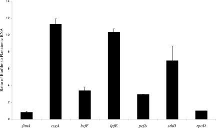
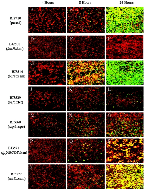
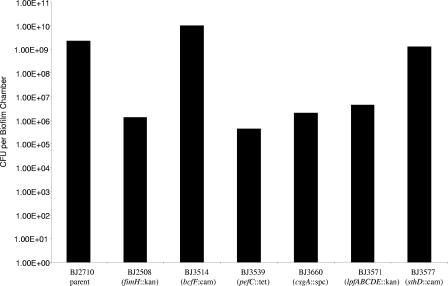
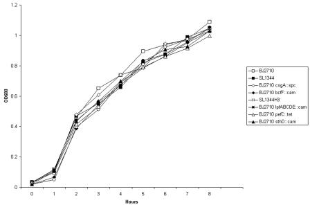
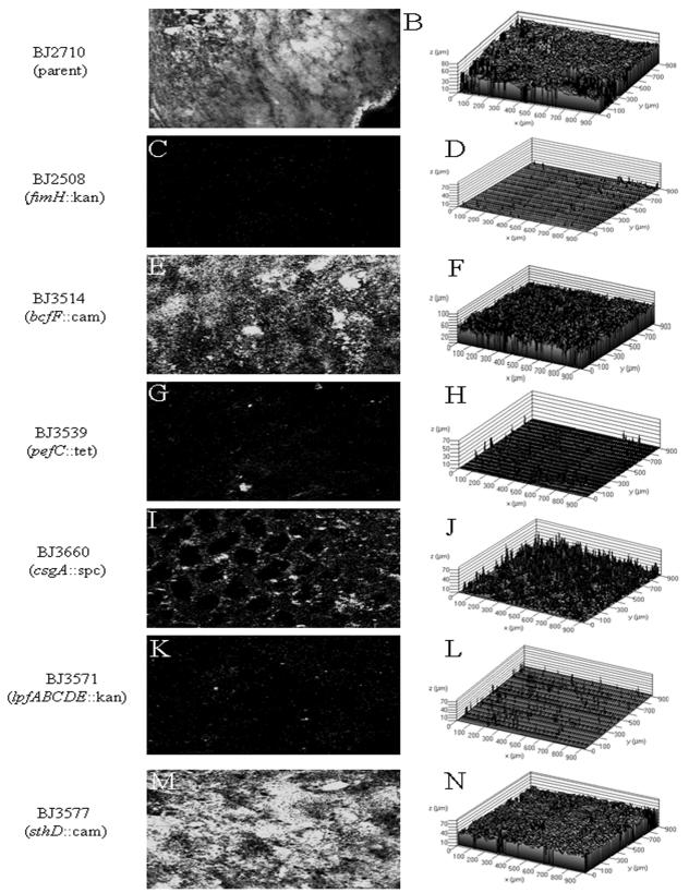
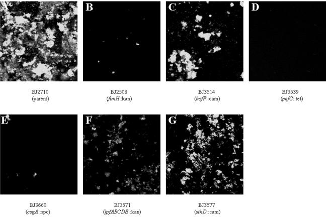
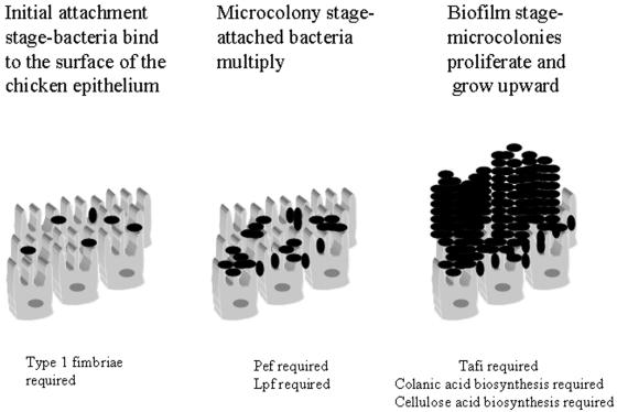
Recent work has demonstrated that Salmonella enterica serovar Typhimurium forms biofilms on HEp-2 tissue culture cells in a type 1 fimbria-dependent manner. To investigate how biofilm growth of HEp-2 tissue culture cells affects gene expression in Salmonella, we compared global gene expression during planktonic growth and biofilm growth. Microarray results indicated that the transcription of approximately 100 genes was substantially altered by growth in a biofilm. These genes encode proteins with a wide range of functions, including antibiotic resistance, central metabolism, conjugation, intracellular survival, membrane transport, regulation, and fimbrial biosynthesis. The identification of five fimbrial gene clusters was of particular interest, as we have demonstrated that type 1 fimbriae are required for biofilm formation on HEp-2 cells and murine intestinal epithelium. Mutations in each of these fimbriae were constructed in S. enterica serovar Typhimurium strain BJ2710, and the mutants were found to have various biofilm phenotypes on plastic, HEp-2 cells, and chicken intestinal tissue. The pef and csg mutants were defective for biofilm formation on each of the three surfaces tested, while the lpf mutant exhibited a complete loss of the ability to form a biofilm on chicken intestinal tissue but only an intermediate loss of the ability to form a biofilm on tissue culture cells and plastic surfaces. The bcf mutant displayed increased biofilm formation on both HEp-2 cells and chicken intestinal epithelium, while the sth mutant had no detectable biofilm defects. In all instances, the mutants could be restored to a wild-type phenotype by a plasmid carrying the functional genes. This is the first work to identify the genomic responses of Salmonella to biofilm formation on host cells, and this work highlights the importance of fimbriae in adhering to and adapting to a eukaryotic cell surface. An understanding of these interactions is likely to provide new insights for intervention strategies in Salmonella colonization and infection.


Figures

FIG. 1.
FIG. 1.
Quantitative RT-PCR comparing fimbrial gene expression in S. enterica serovar Typhimurium strain BJ2710 grown in a 24-h biofilm to that grown planktonically. Total RNA was isolated from either biofilm-grown or planktonically grown BJ2710, converted into cDNA, and used as a template in real-time PCRs. Individual fimbrial genes were quantitated in real time using a TaqMan probe as a transcriptional reporter for each gene examined. The ratio of each fimbrial gene transcript was determined by dividing the biofilm growth expression level by the planktonic growth expression level. The ratio for the control rpoD gene transcript was set at 1 since work in our laboratory has shown that this gene is expressed at constant levels under the two conditions (our unpublished data). The results were averaged and plotted as shown. The P values for the ratios shown for fimA, csgA, bcfF, lpfE, pefA, and sthD are 0.25, 0.01, 0.04, 0.04, 0.07, and 0.02, respectively.
FIG.2.
FIG.2.
Comparison of biofilm formation by S. enterica serovar Typhimurium strains BJ2710 (parent), BJ2508 (BJ2710 fimH::kan), BJ3514 (BJ2710 bcfF::cat), BJ3539 (BJ2710 pefC::tet), BJ3660 (BJ2710 csgA::spc), BJ3571 (BJ2710 lpfABCDE::kan), and BJ3577 (BJ2710 sthD::cat) on cultured HEp-2 cells over a 24-h time course. Shown are composite images of the biofilm formed by adherent S. enterica serovar Typhimurium strain BJ2710 at 4 h (A), 8 h (B), and 24 h (C). Biofilms formed by the indicated Salmonella fimbrial mutants at 4 h (D, G, J, M, P, and S), 8 h (E, H, K, N, Q, and T), and 24 h (F, I, L, O, R, and U) are shown. The bacteria carried pMRP9-1, which encodes GFP, and appeared green under the confocal microscope. The HEp-2 cells were stained with CMTMR and appeared red under the confocal microscope. The level of HEp-2 cell staining for each of the samples was comparable to what is observed in D, which is almost exclusively HEp-2 cell staining.
FIG. 3.
FIG. 3.
Enumeration of bacteria in biofilms formed by S. enterica serovar Typhimurium strains BJ2710 (parent), BJ2508 (BJ2710 fimH::kan), BJ3514 (BJ2710 bcfF::cat), BJ3539 (BJ2710 pefC::tet), BJ3660 (BJ2710 csgA::spc), BJ3571 (BJ2710 lpfABCDE::kan), and BJ3577 (BJ2710 sthD::cat) on HEp-2 cells over 24 h. Biofilms containing bacteria and HEp-2 cells were scraped from the biofilm chambers with a cell scraper, diluted in PBS, and plated onto Lennox agar to determine the number of bacteria in each biofilm. Data collected from three separate biofilm experiments on separate days were averaged and plotted. The data were subjected to statistical analysis using the two-tailed Student's t test. The numbers of organisms isolated from biofilms formed by the fimH, bcfF, pefC, or csgA mutant were found to be significantly different from the numbers of bacteria isolated from the BJ2710 biofilm (P value of >0.05). The difference in the number of organisms isolated from the biofilm formed by BJ2710 and the number of organisms isolated from the biofilm formed by the lpfABCDE mutant was not statistically different, nor was the difference between BJ2710 and the sthD mutant statistically different.
FIG. 4.
FIG. 4.
Growth curves of S. enterica serovar Typhimurium strains BJ2710 (parent), BJ2508 (BJ2710 fimH::kan), BJ3514 (BJ2710 bcfF::cat), BJ3539 (BJ2710 pefC::tet), BJ3660 (BJ2710 csgA::spc), BJ3571 (BJ2710 lpfABCDE::kan), and BJ3577 (BJ2710 sthD::cat) in Lennox broth. Growth curves were performed to ensure that the biofilm phenotypes observed for each fimbrial mutant were not due to a growth defect resulting from the mutation.
FIG. 5.
FIG. 5.
Biofilm formation by S. enterica serovar Typhimurium strains BJ2710 (parent), BJ2508 (BJ2710 fimH::kan), BJ3514 (BJ2710 bcfF::cat), BJ3539 (BJ2710 pefC::tet), BJ3660 (BJ2710 csgA::spc), BJ3571 (BJ2710 lpfABCDE::kan), and BJ3577 (BJ2710 sthD::cat) on chicken intestinal epithelium. Panels A, C, E, G, I, K, and M show x-y-z composite sections generated by confocal microscopy of BJ2710 and its respective fimbrial mutants that were able to adhere to and grow as a biofilm on the chicken intestinal epithelial surface. The organisms were originally labeled with GFP and appear white in the image. Panels B, D, F, H, J, L, and N are topographical maps and correspond to BJ2710 and each of the respective fimbrial mutants and show a dramatic difference in the height and extent of the biofilm formed. The height of biofilm formed varied from 50 to 60 μm (BJ2710, BJ3514, and BJ3577) to less than 10 μm (BJ2508, BJ3539, and BJ3571).
FIG. 6.
FIG. 6.
Biofilm formation by S. enterica serovar Typhimurium strains BJ2710 (wild type), BJ2508 (BJ2710 fimH::kan), BJ3514 (BJ2710 bcfF::cat), BJ3539 (BJ2710 pefC::tet), BJ3660 (BJ2710 csgA::spc), BJ3571 (BJ2710 lpfABCDE::kan), and BJ3577 (BJ2710 sthD::cat) on plastic slides. Images shown in panels A and B depict GFP-labeled BJ2710 and BJ3402, respectively, after growth for 24 h as a biofilm on the plastic slides. Panels C, D, E, F, and G are x-y composite images of strains BJ3514, BJ3539, BJ3660, BJ3571, and BJ3577. Each strain adheres incompletely to the plastic surface; however, each of the fimbrial mutants displayed various levels of adherence to the plastic surface.
FIG. 7.
FIG. 7.
Model of biofilm formation on chicken intestinal epithelium. The first stage in the formation of biofilm is initial attachment. S. enterica serovar Typhimurium strains lacking functional type 1 fimbriae are severely defective in the ability to establish initial colonization of the chicken intestinal epithelium (Fig. 5C). Following initial attachment events, the adherent organisms begin to multiply and form microcolonies on the epithelium. In some cases, the microcolonies can grow to the point that the silhouette of microvilli are visible, as for strain BJ3660, which carries a mutation in the csgA gene (Fig. 5I). A similar phenotype was observed for colanic acid and cellulose biosynthetic genes (37). Mutants in Pef and Lpf are unable to express this biofilm phenotype on chicken intestinal cells and therefore appear to have an early defect in biofilm maturation. Finally, mature biofilm is characterized by almost complete coverage of the epithelial surface and significant height of bacterial growth on top of the mucosa. The S. enterica serovar Typhimurium CsgA mutant as well as colanic acid and cellulose biosynthetic mutants are unable to form mature biofilm, which indicates that those genes are required for the final stages of biofilm formation.

References

    1. Althouse, C., S. Patterson, P. Fedorka-Cray, and R. E. Isaacson. 2003. Type 1 fimbriae of Salmonella enterica serovar Typhimurium bind to enterocytes and contribute to colonization of swine in vivo. Infect. Immun. 71:6446-6452. - PMC - PubMed
    1. Austin, J. W., G. Sanders, W. W. Kay, and S. K. Collinson. 1998. Thin aggregative fimbriae enhance Salmonella enteritidis biofilm formation. FEMS Microbiol. Lett. 162:295-301. - PubMed
    1. Baumler, A. J., and F. Heffron. 1995. Identification and sequence analysis of lpfABCDE, a putative fimbrial operon of Salmonella typhimurium. J. Bacteriol. 177:2087-2097. - PMC - PubMed
    1. Baumler, A. J., R. M. Tsolis, F. A. Bowe, J. G. Kusters, S. Hoffmann, and F. Heffron. 1996. The pef fimbrial operon of Salmonella typhimurium mediates adhesion to murine small intestine and is necessary for fluid accumulation in the infant mouse. Infect. Immun. 64:61-68. - PMC - PubMed
    1. Baumler, A. J., R. M. Tsolis, and F. Heffron. 1996. The lpf fimbrial operon mediates adhesion of Salmonella typhimurium to murine Peyer's patches. Proc. Natl. Acad. Sci. USA 93:279-283. - PMC - PubMed

Publication types

MeSH terms

Substances

LinkOut - more resources