. 2006 Jun;74(6):3180-9.
doi: 10.1128/IAI.02004-05.

Differential requirements for soluble and transmembrane tumor necrosis factor in the immunological control of primary and secondary Listeria monocytogenes infection

Affiliations

Differential requirements for soluble and transmembrane tumor necrosis factor in the immunological control of primary and secondary Listeria monocytogenes infection

Korana Musicki et al. Infect Immun. 2006 Jun.

Abstract

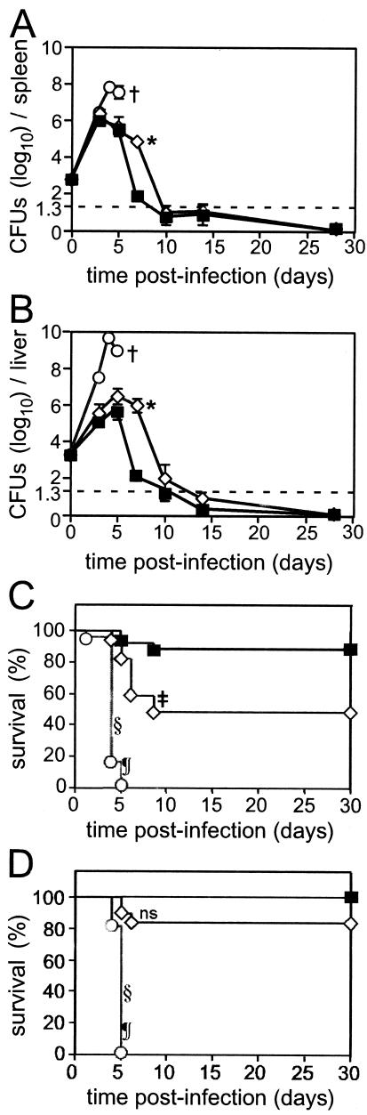
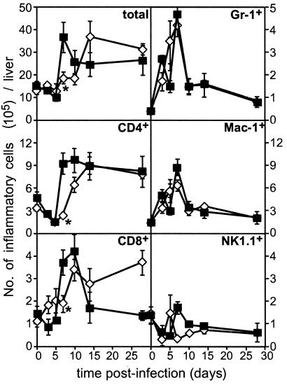
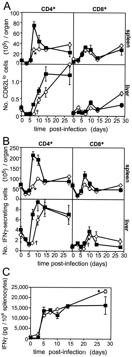
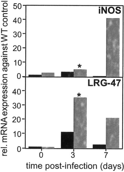
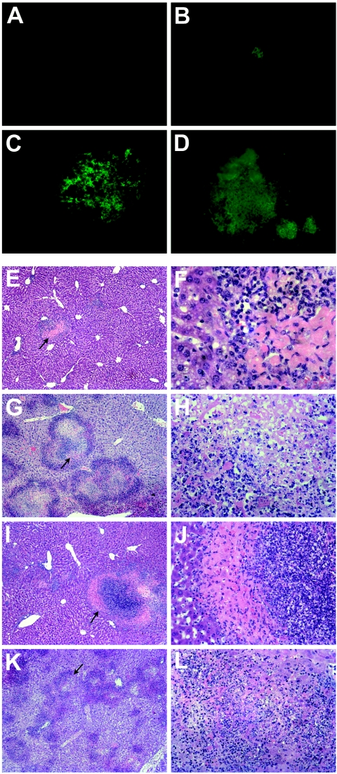
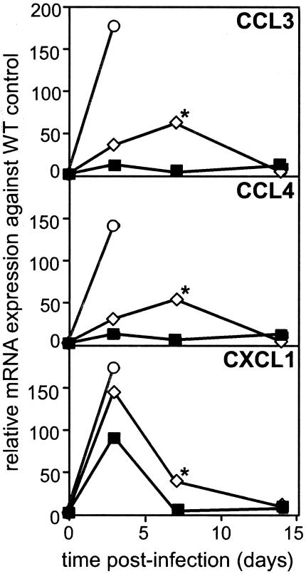
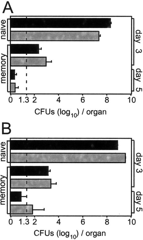
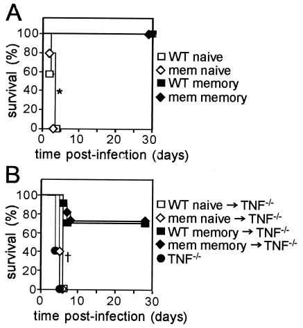
The relative contributions of transmembrane tumor necrosis factor (memTNF) and soluble tumor necrosis factor (solTNF) in innate and adaptive immunity are poorly defined. We examined the capacities of wild-type (WT) mice, TNF-/- mice, and memTNF mice, which express only transmembrane TNF, to control primary and secondary Listeria monocytogenes infections. Soluble TNF was not required for induction or maintenance of protective immunity against a low-dose (200-CFU) Listeria infection. In contrast to TNF-/- mice, both WT and memTNF mice cleared the bacilli within 10 days and were fully protected against rechallenge with a lethal infective dose. Furthermore, T cells transferred from immune mice, but not from naïve, WT, and memTNF mice, protected TNF-/- recipients against an otherwise lethal infection. By contrast, infection with a higher dose of Listeria (2,000 CFU) clearly demonstrated that solTNF is required to coordinate an optimal protective inflammatory response. memTNF mice were more susceptible to a high-dose infection, and they exhibited delayed bacterial clearance, increased inflammation, and necrosis in the liver that resulted in 55% mortality. The dysregulated inflammation was accompanied by prolonged elevated expression of mRNAs for several chemokines as well as the macrophage effector molecules inducible nitric oxide synthase and LRG-47 in the livers of memTNF mice but not in the livers of WT mice. These data demonstrated that memTNF is sufficient for establishing protective immunity against a primary low-dose Listeria infection but that solTNF is required for optimal control of cellular inflammation and resistance to a primary high-dose infection. By contrast, memTNF alone is sufficient for resolution of a secondary, high-dose infection and for the transfer of protective immunity with memory T cells.


Figures

FIG. 1.
FIG. 1.
Delayed bacterial clearance and increased susceptibility of memTNF mice to Listeria infection. WT (▪), memTNF (⋄), and TNF−/− (○) mice were infected with 2,000 (A to C) or 200 (D) CFU of Listeria intravenously. The bacterial loads in the spleen (A) and liver (B) were determined by serial dilution of organ homogenates at different times. The dotted line indicates the limit of detection. The data are the means and standard errors for five mice per group from one of three representative experiments. Significance was determined by ANOVA. An asterisk indicates that the P value was <0.02 for a comparison of memTNF and WT mice, and a dagger indicates that the P value was <0.0001 for a comparison of TNF−/− and WT mice. Additional groups of mice were monitored twice daily and euthanized if they displayed signs of declining health. (C) Time to euthanasia in three experiments following infection of WT (n = 19), memTNF (n = 19), and TNF−/− (n = 11) mice with 2,000 CFU of Listeria. (D) Time to euthanasia in two experiments following infection of WT (n = 21), memTNF (n = 29), and TNF−/− (n = 19) mice with 200 CFU of Listeria. Significance was determined by the log rank Mantel-Cox test. A double dagger indicates that the P value was <0.009 for a comparison of memTNF and WT mice; a section sign indicates that the P value was <0.0001 for a comparison of TNF−/− and WT mice; and a paragraph sign indicates that the P value was 0.0001 for a comparison of memTNF and TNF−/− mice. ns, not significant.
FIG. 2.
FIG. 2.
Delayed influx of T cells in livers of infected memTNF mice. WT (▪) and memTNF (⋄) mice were infected with 2,000 CFU of Listeria intravenously. Livers were perfused, and single-cell suspensions were prepared from uninfected and infected mice at different times. Leukocytes were enumerated, stained for CD4, CD8, Gr-1, Mac-1, and NK1.1, and analyzed by flow cytometry. The data are the means and standard errors for five mice per group from one of two representative experiments. Significance was determined at day 7 by Student's t test. An asterisk indicates that the P value was <0.04 for a comparison of memTNF and WT mice.
FIG. 3.
FIG. 3.
T-lymphocyte activation and IFN-γ secretion in spleens and livers of infected memTNF mice. WT (▪) and memTNF (⋄) mice were infected with 2,000 CFU of Listeria intravenously. (A and B) Single-cell suspensions were prepared from splenocytes and leukocytes from perfused livers of uninfected and infected mice at different times. Leukocytes were enumerated, stained for coexpression of CD4 or CD8 and surface CD62L (A) or intracellular IFN-γ (B), and detected using flow cytometry. (C) Splenocytes were cultured for 72 h with heat-killed L. monocytogenes or medium alone, and IFN-γ production was measured in the culture supernatant by an enzyme-linked immunosorbent assay. The data are the means and standard errors for five mice per group from one of two representative experiments. Significance for the spleen values was determined by ANOVA, and an asterisk indicates that the P value was <0.05 for a comparison of memTNF and WT mice. Significance for the liver values at day 7 was determined by Student's t test, and a dagger indicates that the P value was <0.03 for a comparison of memTNF and WT mice.
FIG. 4.
FIG. 4.
Enhanced macrophage activation in livers of infected memTNF mice. WT (black bars) and memTNF (gray bars) mice were infected with 2,000 CFU of Listeria intravenously. mRNA expression in the livers from uninfected mice and infected mice was measured by RTQ-PCR to determine the relative (rel.) expression of LRG-47 and iNOS. The data are the means and standard errors for five mice per group from one of two representative experiments. Significance was determined by ANOVA. An asterisk indicates that the P value was <0.05 for a comparison of memTNF and WT mice.
FIG. 5.
FIG. 5.
Increased inflammation, necrosis, and iNOS expression in memTNF mice. WT, memTNF, and TNF−/− mice were infected with 2,000 CFU of L. monocytogenes intravenously. The photographs show typical sections from one of five mice in two representative experiments. (A to D) Perfused liver sections were frozen in optimal cutting temperature compound, and 5-μm sections were stained for iNOS expression and examined at a magnification of ×200. (A and B) WT livers on day 3 (A) and day 7 (B). (C and D) memTNF livers on day 3 (C) and day 7 (D). (E to L) Perfused liver sections were fixed in neutral buffered formalin, embedded in paraffin, sectioned, and stained with hematoxylin and eosin. (E, G, I, and K) Magnification, ×25. (F and H) Magnification, ×400. (J and L) Magnification, ×200. (E and F) WT mice 5 days postinfection. (E) Inflammatory foci, scattered throughout the tissue, were mostly discrete and localized and consisted of tightly apposed mononuclear cells. Several necrotic lesions also were evident. (F) The necrotic lesions mostly consisted of a central core of degraded tissue surrounded by neutrophils and mononuclear cells. (G and H) TNF−/− mice 5 days postinfection. (G) Overwhelming hepatic destruction with intense inflammatory involvement was observed throughout the preparation. Darkly staining neutrophils appeared to radiate out from a central region of tissue degradation through the remaining viable liver. (H) High-power view of the interface (arrow in panel G) revealed disintegrated tissue, which was surrounded by cellular debris and pyknotic matter. (I and J) Nonmoribund memTNF mice 5 days postinfection. (I) Inflammatory foci mostly consisted of mononuclear leukocytes and neutrophils, but they were more frequent, larger, less organized, and less compact in memTNF mice than in their WT counterparts. Most necrotic lesions comprised a small area of purulent matter circumscribed by neutrophils and mononuclear cells. Other necrotic lesions consisted of a central core of degraded cells, predominantly neutrophils, surrounded by a region of disintegrated tissue, which was in turn encircled by inflammatory cells. (J) Enlargement (arrow in panel I) showing a central core of dying inflammatory cells and hepatocytes surrounded by completely necrotized tissue and a thin outer layer of leukocytes. (K and L) Moribund memTNF mice 5 days postinfection. (K) Rampant cellular infiltration and hepatic destruction. Darkly staining collections of cells, scattered throughout the tissue, were composed primarily of apoptotic bodies, neutrophils, and necrotic matter. (L) Enlargement of a section (arrow in panel K) showing areas of complete tissue destruction apparent throughout the preparation.
FIG. 6.
FIG. 6.
Enhanced and prolonged chemokine production in livers of infected memTNF mice. WT (▪), memTNF (⋄), and TNF−/− (○) mice were infected with 2,000 CFU of Listeria intravenously. mRNA expression in livers was measured by RTQ-PCR to determine the relative expression of the chemokines CCL3, CCL4, and CXCL1. The data are the means for five mice per group from one of two representative experiments. The significance of differences at day 7 was determined by Student's t test. An asterisk indicates that the P value was <0.04 for a comparison of memTNF and WT mice.
FIG. 7.
FIG. 7.
Transmembrane TNF is sufficient to control bacterial growth following a secondary challenge with a lethal dose of Listeria. WT (black bars) and memTNF (gray bars) mice were infected with 200 CFU of Listeria intravenously. Eight weeks after the initial infection, naïve and immune mice were infected with 105 CFU of Listeria. The bacterial loads in the spleen (A) and liver (B) were determined at 3 and 5 days postinfection. The dotted line indicates the limit of detection. The data are the means and standard errors for five mice per group from one of two representative experiments.
FIG. 8.
FIG. 8.
Protection against secondary Listeria infection does not require soluble TNF. (A) WT and memTNF mice were infected with 200 CFU of Listeria intravenously, and 8 weeks after the initial infection, naïve and immune mice were infected with 105 CFU of Listeria. The data are the time to euthanasia for between 8 and 10 mice/group. Significance was determined by the log rank Mantel-Cox test. The asterisk indicates that the P value was <0.001 for a comparison of immune and naïve mice. (B) WT and memTNF mice were infected with 200 CFU of Listeria intravenously, and 14 days later, single-spleen-cell suspensions were prepared from immune and naïve mice, and T cells were isolated by negative selection (CD3+, >90%). Two hundred colony-forming units of Listeria and 5 × 10⁶ purified T cells were injected intravenously into TNF−/− mice that had been irradiated 24 h previously with 500 rads. The data are the time to euthanasia for 11 to 15 mice/group. Significance was determined by the log rank Mantel-Cox test. The dagger indicates that the P value was <0.002 for comparisons of WT immune and WT naïve mice and memTNF immune and memTNF naïvemice.

References

    1. Bouwer, H. G., R. A. Barry, and D. J. Hinrichs. 1997. Acquired immunity to an intracellular pathogen: immunologic recognition of L. monocytogenes-infected cells. Immunol. Rev. 158:137-146. - PubMed
    1. Bowie, V. L., K. A. Snella, A. S. Gopalachar, and P. Bharadwaj. 2004. Listeria meningitis associated with infliximab. Ann. Pharmacother. 38:58-61. - PubMed
    1. Britton, W. J., N. Meadows, D. A. Rathjen, D. R. Roach, and H. Briscoe. 1998. A tumor necrosis factor mimetic peptide activates a murine macrophage cell line to inhibit mycobacterial growth in a nitric oxide-dependent fashion. Infect. Immun. 66:2122-2127. - PMC - PubMed
    1. Collazo, C. M., G. S. Yap, G. D. Sempowski, K. C. Lusby, L. Tessarollo, G. F. Woude, A. Sher, and G. A. Taylor. 2001. Inactivation of LRG-47 and IRG-47 reveals a family of interferon gamma-inducible genes with essential, pathogen-specific roles in resistance to infection. J. Exp. Med. 194:181-188. - PMC - PubMed
    1. Czuprynski, C. J., J. F. Brown, N. Maroushek, R. D. Wagner, and H. Steinberg. 1994. Administration of anti-granulocyte mAb RB6-8C5 impairs the resistance of mice to Listeria monocytogenes infection. J. Immunol. 152:1836-1846. - PubMed

Publication types

MeSH terms