. 2006 Jun;74(6):3488-97.
doi: 10.1128/IAI.02006-05.

The major subunit, CfaB, of colonization factor antigen i from enterotoxigenic Escherichia coli is a glycosphingolipid binding protein

Affiliations

The major subunit, CfaB, of colonization factor antigen i from enterotoxigenic Escherichia coli is a glycosphingolipid binding protein

Lena Jansson et al. Infect Immun. 2006 Jun.

Abstract

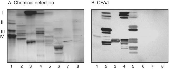
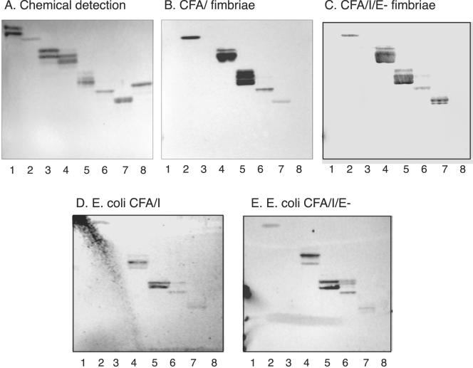
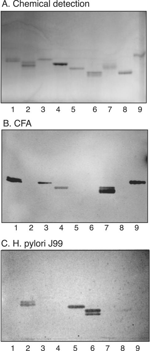
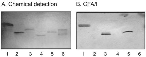
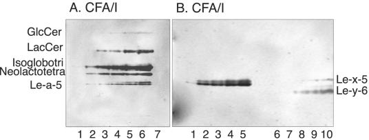
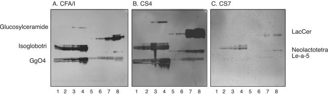
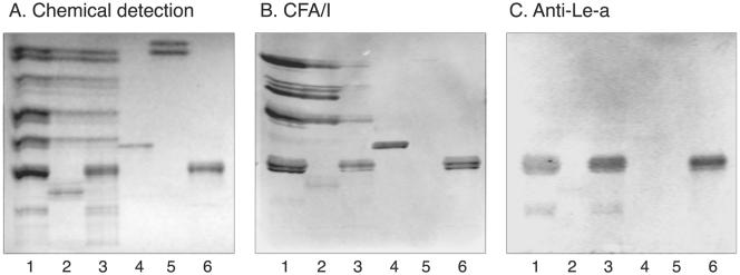
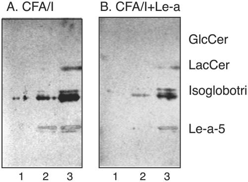
Bacterial adherence to mucosal surfaces is an important virulence trait of pathogenic bacteria. Adhesion of enterotoxigenic Escherichia coli (ETEC) to the intestine is mediated by a number of antigenically distinct colonization factors (CFs). One of the most common CFs is CFA/I. This has a fimbrial structure composed of a major repeating subunit, CfaB, and a single tip subunit, CfaE. The potential carbohydrate recognition by CFA/I was investigated by binding CFA/I-fimbriated bacteria and purified CFA/I fimbriae to a large number of variant glycosphingolipids separated on thin-layer chromatograms. For both fimbriated bacteria and purified fimbriae, specific interactions could be identified with a number of nonacid glycosphingolipids. These included glucosylceramide, lactosylceramide with phytosphingosine and/or hydroxy fatty acids, neolactotetraosylceramide, gangliotriaosylceramide, gangliotetraosylceramide, the H5 type 2 pentaglycosylceramide, the Lea-5 glycosphingolipid, the Lex-5 glycosphingolipid, and the Ley-6 glycosphingolipid. These glycosphingolipids were also recognized by recombinant E. coli expressing CFA/I in the absence of tip protein CfaE, as well as by purified fimbriae from the same strain. This demonstrates that the glycosphingolipid-binding capacity of CFA/I resides in the major CfaB subunit.


Figures

FIG. 1.
FIG. 1.
Binding of 125I-labeled CFA/I fimbriae to mixtures of glycosphingolipids on thin-layer chromatograms. Chemical detection by anisaldehyde (A) and an autoradiogram obtained by binding of 125I-labeled CFA/I fimbriae (B) are shown. The glycosphingolipids were separated on aluminum-backed silica gel plates with chloroform-methanol-water (60:35:8, by volume) as the solvent system, and the binding assay was performed as described in Materials and Methods. Lanes: 1, nonacid glycosphingolipids of human blood group A erythrocytes (40 μg); 2, nonacid glycosphingolipids of guinea pig intestine (40 μg); 3, nonacid glycosphingolipids of mouse feces (40 μg); 4, nonacid glycosphingolipids of rat intestine (40 μg); 5, nonacid glycosphingolipids of human meconium (40 μg); 6, calf brain gangliosides (40 μg); 7, acid glycosphingolipids of human erythrocytes (40 μg); 8, acid glycosphingolipids of human hypernephroma (40 μg). Autoradiography was performed for 12 h. The roman numerals to the left indicate the approximate numbers of carbohydrate residues in the bands.
FIG. 2.
FIG. 2.
Binding of CFA/I fimbriae, CFA/I/E fimbriae, and recombinant bacterial cells expressing CFA/I fimbriae and CFA/I/E fimbriae to pure glycosphingolipids on thin-layer chromatograms. Shown are chemical detection by anisaldehyde (A) and autoradiograms obtained by binding of CFA/I fimbriae (B), CFA/I/E fimbriae (C), CFA/I-expressing E. coli (strain Top10-CFA/I) (D), and E. coli with CFA/I/E fimbriae (strain Top10-CFA/I/E) (E). The glycosphingolipids were separated on aluminum-backed silica gel plates with chloroform-methanol-water (60:35:8, by volume) as the solvent system, and the binding assays were performed as described in Materials and Methods. Autoradiography was performed for 12 h. Lanes: 1, galactosylceramide (Galβ1Cer) (2 μg); 2, glucosylceramide (Glcβ1Cer) (2 μg); 3, lactosylceramide (Galβ4Glcβ1Cer) with d18:1-16:0-24:0 (2 μg); 4, lactosylceramide (Galβ4Glcβ1Cer) with t18:0-h16:0-h24:0 (2 μg); 5, isoglobotriaosylceramide (Galα3Galβ4Glcβ1Cer) (2 μg); 6, neolactotetraosylceramide (Galβ4GlcNAcβ3Galβ4Glcβ1Cer) (2 μg); 7, Lea pentaglycosylceramide [Galβ3(Fucα4)GlcNAcβ3Galβ4Glcβ1Cer] (2 μg); 8, globotetraosylceramide (GalNAcβ3Galα4Galβ4Glcβ1Cer) (2 μg).
FIG. 3.
FIG. 3.
Comparison of glycosphingolipid recognition by CFA/I fimbriae of ETEC and BabA-expressing H. pylori. The glycosphingolipids were chromatographed on aluminum-backed silica gel plates and visualized with anisaldehyde (A). Duplicate chromatograms were incubated with 125I-labeled CFA/I fimbriae (B) and 35S-labeled H. pylori strain J99 (C), followed by autoradiography for 12 h, as described in Materials and Methods. The solvent system used was chloroform-methanol-water (60:35:8, by volume). Lanes: 1, Lea pentaglycosylceramide [Galβ3(Fucα4)GlcNAcβ3Galβ4Glcβ1Cer] (2 μg); 2, B type 1 hexaglycosylceramide [Galα3(Fucα2)Galβ3GlcNAcβ3Galβ4Glcβ1Cer] (2 μg); 3, Lex pentaglycosylceramide [Galβ4(Fucα3)GlcNAcβ3Galβ4 Glcβ1Cer] (2 μg); 4, B hepta-glycosylceramide (Galα3Galβ4GlcNAcβ3Galβ4GlcNAcβ3Galβ4Glcβ1Cer) (2 μg); 5, Leb hexaglycosylceramide [Fucα2Galβ3(Fucα4)GlcNAcβ3Galβ4Glcβ1Cer] (2 μg); 6, A type 1 heptaglycosylceramide [GalNAcα3(Fucα2)Galβ3(Fucα4)GlcNAcβ3Galβ4Glcβ1Cer] (2 μg); 7, Ley hexaglycosylceramide [Fucα2Galβ4(Fucα3)GlcNAcβ3Galβ4Glcβ1Cer] (2 μg); 8, A type 2 heptaglycosylceramide [GalNAcα3(Fucα2)Galβ4(Fucα3)GlcNAcβ3Galβ4Glcβ1Cer] (2 μg); 9, H type 2 pentaglycosylceramide (Fucα2Galβ4GlcNAcβ3Galβ4Glcβ1Cer) (2 μg). Autoradiography was performed for 12 h.
FIG. 4.
FIG. 4.
Binding of CFA-1 fimbriae of ETEC to pure glycosphingolipids on thin-layer chromatograms. Chemical detection by anisaldehyde (A) and an autoradiogram obtained by binding of 125I-labeled CFA-1 fimbriae (B) are shown. The glycosphingolipids were separated on aluminum-backed silica gel plates with chloroform-methanol-water (60:35:8, by volume) as the solvent system, and the binding assays were performed as described in Materials and Methods. Autoradiography was performed for 12 h. Lanes: 1, neolactotetraosylceramide (Galβ4GlcNAcβ3Galβ4Glcβ1Cer) (2 μg); 2, sialyl-neolactotetraosylceramide (NeuAcα3Galβ4GlcNAcβ3Galβ4Glcβ1Cer) (2 μg); 3, Lex pentaglycosylceramide [Galβ4(Fucα3)GlcNAcβ3Galβ4Glcβ1Cer] (2 μg); 4, sialyl-Lex hexaglycosylceramide [NeuAcα3Galβ4(Fucα3)GlcNAcβ3Galβ4Glcβ1Cer] (2 μg); 5, Lea pentaglycosylceramide [Galβ3(Fucα4)GlcNAcβ3Galβ4Glcβ1Cer] (2 μg); 6, sialyl-Lea hexaglycosylceramide [NeuAcα3Galβ3(Fucα4) GlcNAcβ3Galβ4Glcβ1Cer] (2 μg).
FIG. 5.
FIG. 5.
Binding of CFA/I fimbriae of ETEC to serial dilutions of glycosphingolipids. Autoradiograms were obtained by binding of 125I-labeled CFA/I fimbriae to serial dilutions (0.1 to 1.0 μg) of glycosphingolipids in a chromatogram binding assay. The binding assay was done as described in Materials and Methods. Autoradiography was performed for 12 h. Lanes in panel A: 1 to 6, glucosylceramide (Glcβ1Cer), lactosylceramide (Galβ4Glcβ1Cer) with t18:0-h16:0-h24:0, isoglobotriaosylceramide (Galα3Galβ4Glcβ1Cer), neolactotetraosylceramide (Galβ4GlcNAcβ3Galβ4Glcβ1Cer), and Lea pentaglycosylceramide [Galβ3(Fucα4)GlcNAcβ3Galβ4Glcβ1Cer] (0.1 to 1.0 μg of each compound); 7, negative control globotetraosylceramide (GalNAcβ3Galα4Galβ4Glcβ1Cer) (4 μg). Lanes in panel B: 1 to 5, Lea pentaglycosylceramide [Galβ3(Fucα4)GlcNAcβ3Galβ4Glcβ1Cer] (0.1 to 0.8 μg); 6 to 10, Lex pentaglycosylceramide [Galβ4(Fucα3)GlcNAcβ3Galβ4Glcβ1Cer] and Ley hexaglycosylceramide [Fucα2Galβ4(Fucα3)GlcNAcβ3Galβ4Glcβ1Cer] (0.1 to 0.8 μg of each compound).
FIG. 6.
FIG. 6.
Electron micrograph of immunolabeled and negatively stained recombinant E. coli expressing CFA/I fimbriae without tip protein CfaE (strain Top10-CFA/I/E). The bacteria were labeled with monoclonal anti-CFA/I antibody 1:6. The bar represents 1 μm.
FIG. 7.
FIG. 7.
Comparison of glycosphingolipid recognition of CFA/I and heterologous CF fimbriae of ETEC. Autoradiograms were obtained by binding of 125I-labeled CFA/I fimbriae (A), CS4 fimbriae (B), and CS7 fimbriae (C) to serial dilutions (0.4 to 2.0 μg) of glycosphingolipids in a chromatogram binding assay. The binding assay was done as described in Materials and Methods. The solvent system used was chloroform-methanol-water (60:35:8, by volume). Autoradiography was performed for 12 h. Lanes: 1 to 4, glucosylceramide (Glcβ1Cer), isoglobotriaosylceramide (Galα3Galβ4Glcβ1Cer), and gangliotetraosylceramide (Galβ3GalNAcβ4Galβ4Glcβ1Cer) (0.4 to 2.0 μg of each compound); 5 to 8, lactosylceramide (Galβ4Glcβ1Cer) with t18:0-h16:0-h24:0, neolactotetraosylceramide (Galβ4GlcNAcβ3Galβ4Glcβ1Cer), and Lea pentaglycosylceramide [Galβ3(Fucα4)GlcNAcβ3Galβ4Glcβ1Cer] (0.4 to 2.0 μg of each compound).
FIG. 8.
FIG. 8.
Binding of CFA/I fimbriae of ETEC to nonacid glycosphingolipids of human small intestine. The glycosphingolipids were separated on aluminum-backed silica gel plates and visualized with anisaldehyde (A). Duplicate chromatograms were incubated with 125I-labeled CFA/I fimbriae (B) and monoclonal antibodies directed against the Lea determinant (C), followed by autoradiography for 12 h, as described in Materials and Methods. The solvent system used was chloroform-methanol-water (60:35:8, by volume). Lanes: 1 to 3, nonacid glycosphingolipids of human small intestine of three different individuals (40 μg/lane); 4, reference neolactotetraosylceramide (Galβ4GlcNAcβ3Galβ4Glcβ1Cer) (4 μg); 5, reference galactosylceramide (Galβ1Cer) (4 μg); 6, reference Lea pentaglycosylceramide [Galβ3(Fucα4)GlcNAcβ3Galβ4Glcβ1Cer] (4 μg).
FIG. 9.
FIG. 9.
Effect of preincubation of CFA/I fimbriae with oligosaccharides. Radiolabeled CFA/I fimbriae were incubated with Lea pentasaccharide (1 mg/ml) in PBS for 2 h at room temperature. The suspensions were then utilized in a chromatogram binding assay. Panels: A, binding of CFA/I fimbriae alone; B, binding of CFA/I fimbriae incubated with Lea pentasaccharide. The lanes contained dilutions of glucosylceramide (Glcβ1Cer), lactosylceramide with hydroxy ceramide (Galβ4Glcβ1Cer), isoglobotriaosylceramide (Galα3Galβ4Glcβ1Cer), and Lea pentaglycosylceramide [Galβ3(Fucα4)GlcNAcβ3Galβ4Glcβ1Cer] (0.1 to 0.4 μg of each compound). The glycosphingolipids were separated on aluminum-backed silica gel plates with chloroform-methanol-water (60:35:8, by volume) as the solvent system, and the binding assay was performed as described in Materials and Methods. Autoradiography was performed for 12 h.

References

    1. Anantha, R. P., A. L. McVeigh, L. H. Lee, M. K. Agnew, F. J. Cassels, D. A. Scott, T. S. Whittam, and S. J. Savarino. 2004. Evolutionary and functional relationships of colonization factor antigen I and other class 5 adhesive fimbriae of enterotoxigenic Escherichia coli. Infect. Immun. 72:7190-7201. - PMC - PubMed
    1. Ångström, J., S. Teneberg, M. Abul Milh, T. Larsson, I. Leonardsson, B.-M. Olsson, M. Ölwegård Halvarsson, D. Danielsson, I. Näslund, Å. Ljungh, T. Wadström, and K.-A. Karlsson. 1998. The lactosylceramide binding specificity of Helicobacter pylori. Glycobiology 8:297-309. - PubMed
    1. Aspholm-Hurtig, M., G. Dailide, M. Lahmann, A. Kalia, D. Ilver, N. Roche, S. Vikström, R. Sjöström, S. Lindén, A. Bäckström, A. Arnqvist, J. Mahdavi, U. J. Nilsson, B. Velapatiño, R. H. Gilman, M. Gerhard, T. Alarcon, M. López-Brea, T. Nakazawa, J. G. Fox, P. Correa, M. G. Dominguez-Bello, G. I. Perez-Perez, M. J. Blaser, S. Normark, I. Carlstedt, S. Oscarson, S. Teneberg, D. E. Berg, and T. Borén. 2004. Functional adaptation of BabA, the Helicobacter pylori blood-group antigen binding adhesin. Science 305:519-522. - PubMed
    1. Bartus, H., P. Actor, E. Snipes, D. Sedlock, and I. Zajac. 1985. Indications that the erythrocyte receptor involved in enterotoxigenic Escherichia coli attachment is a sialoglycoconjugate. J. Clin. Microbiol. 21:951-954. - PMC - PubMed
    1. Björk, S., M. E. Breimer, G. C. Hansson, K.-A. Karlsson, and H. Leffler. 1987. Structures of blood group glycosphingolipids in human small intestine. A relation between the expression of fucolipids of epithelial cells and the ABO, Le and Se phenotype of the donor. J. Biol. Chem. 262:6758-6765. - PubMed

Publication types

MeSH terms

LinkOut - more resources