. 2006 Jun;74(6):3643-50.
doi: 10.1128/IAI.00012-06.

Infection of human fallopian tube epithelial cells with Neisseria gonorrhoeae protects cells from tumor necrosis factor alpha-induced apoptosis

Affiliations

Infection of human fallopian tube epithelial cells with Neisseria gonorrhoeae protects cells from tumor necrosis factor alpha-induced apoptosis

Priscilla Morales et al. Infect Immun. 2006 Jun.

Abstract

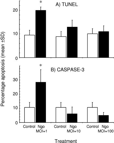
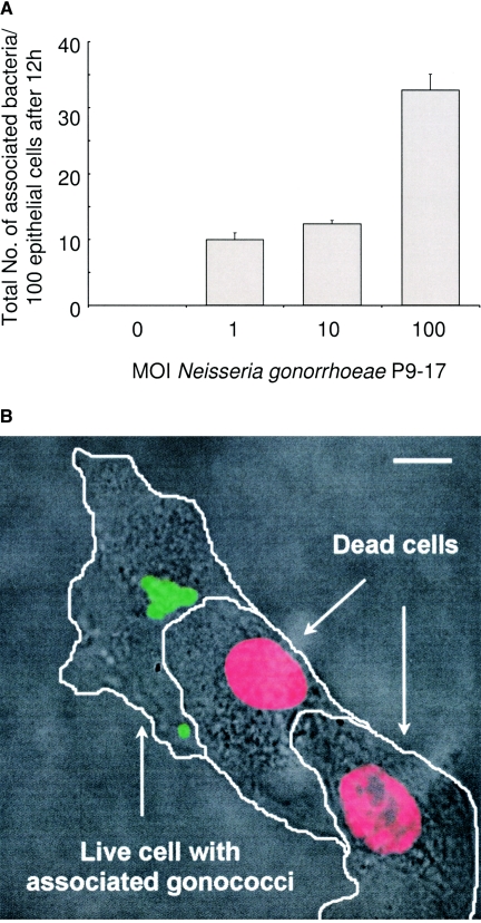
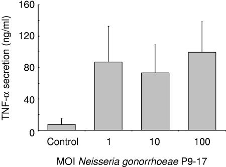
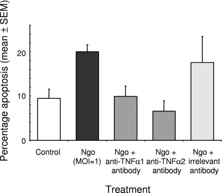
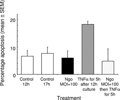
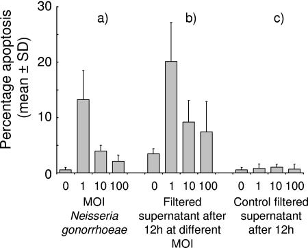
Following infection with Neisseria gonorrhoeae, bacteria may ascend into the Fallopian tubes (FT) and induce salpingitis, a major cause of infertility. In the FT, interactions between mucosal epithelial cells and gonococci are pivotal events in the pathogen's infection cycle and the inflammatory response. In the current study, primary FT epithelial cells were infected in vitro with different multiplicities of infection (MOI) of Pil+ Opa+ gonococci. Bacteria showed a dose-dependent association with cells and induced the secretion of tumor necrosis factor alpha (TNF-alpha). A significant finding was that gonococcal infection (MOI = 1) induced apoptosis in approximately 30% of cells, whereas increasing numbers of bacteria (MOI = 10 to 100) did not induce apoptosis. Apoptosis was observed in only 11% of cells with associated bacteria, whereas >84% of cells with no adherent bacteria were apoptotic. TNF-alpha was a key contributor to apoptosis, since (i) culture supernatants from cells infected with gonococci (MOI = 1) induced apoptosis in naïve cultures, suggesting that a soluble factor was responsible; (ii) gonococcal infection-induced apoptosis was inhibited with anti-TNF-alpha antibodies; and (iii) the addition of exogenous TNF-alpha induced apoptosis, which was inhibited by the presence of increasing numbers of bacteria (MOI = 10 to 100). These data suggest that TNF-alpha-mediated apoptosis of FT epithelial cells is likely a primary host defense mechanism to prevent pathogen colonization. However, epithelial cell-associated gonococci have evolved a mechanism to protect the cells from undergoing TNF-alpha-mediated apoptosis, and this modulation of the host innate response may contribute to establishment of infection. Understanding the antiapoptotic mechanisms used by Neisseria gonorrhoeae will inform the pathogenesis of salpingitis and could suggest new intervention strategies for prevention and treatment of the disease.


Figures

FIG. 1.
FIG. 1.
Apoptosis in human Fallopian tube epithelial cells challenged in vitro with Neisseria gonorrhoeae. Apoptosis was detected in cultures of primary human FT epithelial cells 12 h after challenge with Neisseria gonorrhoeae (Ngo; MOI = 1, 10, or 100) by (A) TUNEL assay and (B) detection of caspase-3 activity. Each column denotes the mean and SEM (error bar) from 10 experiments (MOI = 1), 8 experiments (MOI = 10), or 7 experiments (MOI = 100) carried out with FT epithelial cells from 5 and 10 donors for the caspase-3 and TUNEL assays, respectively.
FIG. 2.
FIG. 2.
(A) Association of Neisseria gonorrhoeae with FT epithelial cells, determined after a 12-h challenge with different MOIs of bacteria by confocal microscopy. Each column denotes the mean association and SEM (error bar) from three independent experiments. (B) Association of Neisseria gonorrhoeae protects human FT epithelial cells from apoptosis. Cultures of primary human FT epithelial cells were challenged with Neisseria gonorrhoeae (MOI = 1) for 12 h, and the confocal image, which is representative of 10 experiments carried out with FT from 10 donors, clearly shows the apoptotic phenotype (red-nucleated cells, determined by TUNEL staining) present in two cells without associated bacteria, whereas the single cell with associated bacteria is viable. Bar, 10 μm.
FIG. 3.
FIG. 3.
Secretion of TNF-α from FT epithelial cells following challenge with different MOIs of Neisseria gonorrhoeae for 12 h. Each column represents the mean TNF-α level and SEM (error bar) from experiments with cells from five individual donors.
FIG. 4.
FIG. 4.
Correlation between apoptosis of FT epithelial cells induced by Neisseria gonorrhoeae and the presence of TNF-α. Cultures of primary human FT epithelial cells were challenged with Neisseria gonorrhoeae (Ngo; MOI = 1) for 12 h in the presence and absence of anti-human TNF-α1 and TNF-α2 antibodies. Apoptosis induction was detected by TUNEL assay and analyzed by confocal microscopy. As a control, cells were also challenged with Neisseria gonorrhoeae in the presence of an irrelevant antibody. Data are from six experiments carried out with FT epithelial cells from six different donors, with the columns representing the means and the error bars representing the SEM.
FIG. 5.
FIG. 5.
Inhibition of TNF-α-induced apoptosis in FT epithelial cells by Neisseria gonorrhoeae. Apoptosis in cultures of primary human FT epithelial cells was induced by treatment with exogenous TNF-α for 5 h. Challenge with Neisseria gonorrhoeae (Ngo; MOI = 100) for 12 h did not induce apoptosis. Cells challenged with gonococci before treatment with exogenous TNF-α were protected from the proapoptotic effects of the cytokine. Data are from six experiments carried out with FT epithelial cells from six different donors, with the columns representing the means and the error bars representing the SEM.
FIG. 6.
FIG. 6.
Effects of supernatants from FT epithelial cell cultures infected for 12 h with gonococci on naïve cell cultures. Culture supernatants from FT epithelial cells that had been infected with gonococci (MOI = 1, 10, or 100) for 12 h were centrifuged to remove bacteria, filtered (0.2-μm pore size), and applied to fresh FT epithelial cells, and apoptosis was then determined by TUNEL assay following a further 12-h incubation. (a) Apoptosis induced by gonococci after the usual 12-h challenge. (b) Apoptosis of naïve FT epithelial cells induced by supernatants taken from cell cultures infected with different MOIs for 12 h. (c) Apoptosis of naïve FT epithelial cells induced by supernatants taken from control, uninfected cell cultures. Each column represents the mean and SEM (error bar) of nine experiments with cells from three donors.

Similar articles

Cited by

References

    1. Ayala, P., J. Scott Wilbur, L. M. Wetzler, J. A. Tainer, A. Snyder, and M. So. 2005. The pilus and porin of Neisseria gonorrhoeae cooperatively induce Ca2+ transients in infected epithelial cells. Cell. Microbiol. 7:1736-1748. - PubMed
    1. Beck, S. C., and T. F. Meyer. 2000. IgA1 protease from Neisseria gonorrhoeae inhibits TNF alpha-mediated apoptosis of human monocytic cells. FEBS Lett. 472:287-292. - PubMed
    1. Binnicker, M. J., R. D. Williams, and M. A. Apicella. 2003. Infection of human urethral epithelium with Neisseria gonorrhoeae elicits an upregulation of host anti-apoptotic factors and protects cells from staurosporine-induced apoptosis. Cell. Microbiol. 5:549-560. - PubMed
    1. Binnicker, M. J., R. D. Williams, and M. A. Apicella. 2004. Gonococcal porin IB activates NF-kappa B in human urethral epithelium and increases the expression of host antiapoptotic factors. Infect. Immun. 72:6408-6417. - PMC - PubMed
    1. Brunham, R. C., B. Binns, F. Guijon, D. Danforth, M. L. Kosseim, F. Rand, J. Mcdowell, and E. Rayner. 1988. Etiology and outcome of acute pelvic inflammatory disease. J. Infect. Dis. 158:510-517. - PubMed

Publication types

Substances

LinkOut - more resources