Review
. 2006 Jun;7(6):423-36.
doi: 10.1038/nrn1928.

Purinergic signalling in neuron-glia interactions

Affiliations
Review

Purinergic signalling in neuron-glia interactions

R Douglas Fields et al. Nat Rev Neurosci. 2006 Jun.

Abstract

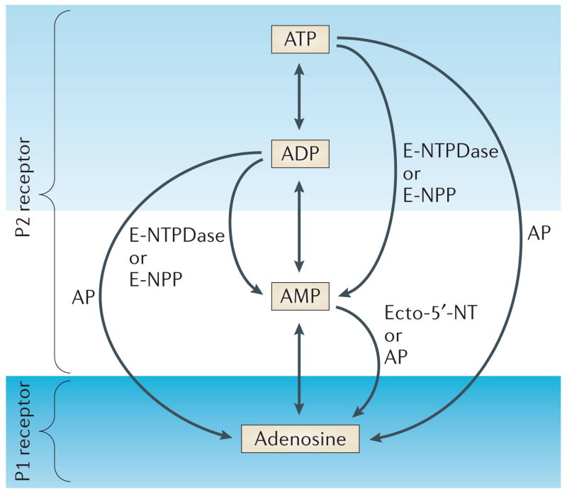
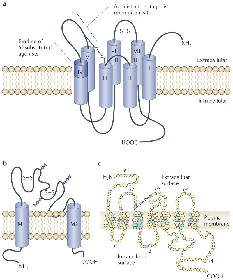
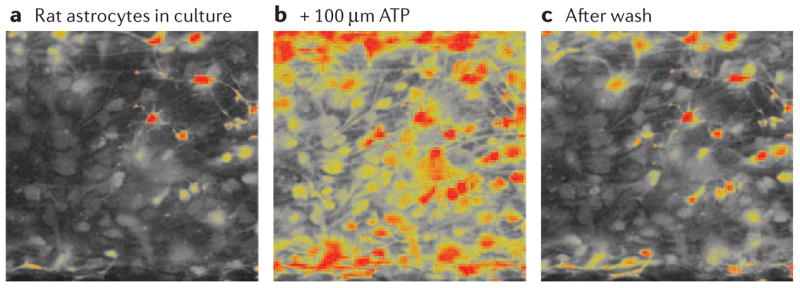
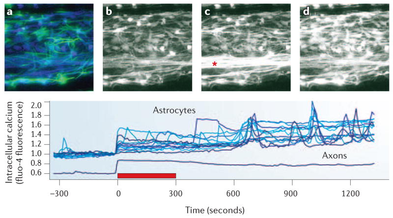
Activity-dependent release of ATP from synapses, axons and glia activates purinergic membrane receptors that modulate intracellular calcium and cyclic AMP. This enables glia to detect neural activity and communicate among other glial cells by releasing ATP through membrane channels and vesicles. Through purinergic signalling, impulse activity regulates glial proliferation, motility, survival, differentiation and myelination, and facilitates interactions between neurons, and vascular and immune system cells. Interactions among purinergic, growth factor and cytokine signalling regulate synaptic strength, development and responses to injury. We review the involvement of ATP and adenosine receptors in neuron-glia signalling, including the release and hydrolysis of ATP, how the receptors signal, the pharmacological tools used to study them, and their functional significance.


Figures

Figure 1
Figure 1. Purinergic receptors bind extracellular ATP and the reaction products that result from its enzymatic hydrolysis by ectonucleotidases
P2 receptors bind ATP and ADP, whereas P1 receptors bind adenosine. The metabolism of extracellular ATP is regulated by several ectonucleotidases, including members of the E-NTPDase (ectonucleoside triphosphate diphosphohydrolase) family and the E-NPP (ectonucleotide pyrophosphatase/phosphodiesterase) family. Ecto-5′-nucleotidase (Ecto-5′-NT) and alkaline phosphatase (AP) catalyse the nucleotide degradation to adenosine.
Figure 2
Figure 2. Membrane receptors for extracellular ATP and adenosine
The P1 family of receptors for extracellular adenosine are G-protein-coupled receptors that signal by inhibiting or activating adenylate cyclase (a). The P2 family of receptors bind extracellular ATP or ADP, and are comprised of two types of receptor (P2X and P2Y). The P2X family of receptors are ligand-gated ion channels (b) and the P2Y family are G-protein-coupled receptors (c). S–S, disulphide bond; e1–e4, extracellular domain loops 1–4; i1–i4, intracellular domain loops 1–4. Panel a modified, with permission, from REF. © (1998) Elsevier Science. Panel b reproduced, with permission, from REF. © (1994) Macmillan Publishers Ltd. Panel c modified, with permission, from REF. © (2006) Elsevier Science.
Figure 3
Figure 3. Astrocytes have several types of ionotropic and metabotropic membrane receptor for ATP and its breakdown products, ADP, AMP and adenosine, which increase intracellular calcium concentrations
Calcium transients and waves induced by extracellular ATP are important in intercellular communication between astrocytes and between neurons and astrocytes. a | Rat astrocytes in culture. b | Application of 100 μM ATP to cultured rat astrocytes resulted in rapid and large increases in intracellular calcium concentrations. c | Shows recovery after a 15 min wash. Intracellular calcium concentration was monitored using confocal microscopy to measure the fluorescence intensity of the calcium-sensitive indicator, fluo-4. High concentrations of intracellular calcium are indicated in yellow and orange.
Figure 4
Figure 4. Calcium imaging reveals that glial cells can respond to electrical activity in axons
Impulse activity causes the release of ATP and other neurotransmitters from synapses and axons. This, in turn, activates membrane receptors on several types of glia, triggering increases in intracellular calcium concentration. Intracellular calcium responses in rat astrocytes are shown in response to 10 Hz of electrical stimulation of dorsal root ganglion axons (*) in co-culture. Increasing concentrations of intracellular calcium are indicated by increased fluorescence intensity of the calcium indicator, fluo-4. a | Immunocytochemical staining for glial fibrillary acidic protein (GFAP, green), a cytoskeletal protein expressed in astrocytes, is shown superimposed on the same field used for live-cell calcium imaging (blue). Not all astrocytes express GFAP at high levels at this stage (48 h in co-culture). bd | Represent calcium imaging taken from cells at −300 s (b), 20 s (c) and 630 s (d) with respect to the 5 min train of 10 Hz action potentials (red bar in plot). The changes in the intracellular calcium concentrations in astrocytes as a result of neuronal firing are shown in the graph (bottom panel). There are no synapses in these cultures. For methods, see REF. . Note that intracellular calcium values in axons have been displaced by 0.4 to better distinguish axon responses from astrocyte responses in the plot. See also online Supplementary information S1 (movie).

References

    1. Altevogt BM, Paul DL. Four classes of intercellular channels between glial cells in the CNS. J Neurosci. 2004;24:4313–4323. - PMC - PubMed
    1. Lev-Ram V, Ellisman MH. Axonal activation-induced calcium transients in myelinating Schwann cells, sources, and mechanisms. J Neurosci. 1995;15:2628–2637. - PMC - PubMed
    1. McCarthy KD, Salm AK. Pharmacologically-distinct subsets of astroglia can be indentified by their calcium response to neuroligands. Neuroscience. 1991;41:325–333. - PubMed
    1. Porter JT, McCarthy KD. Hippocampal astrocytes in situ respond to glutamate released from synaptic terminals. J Neurosci. 1996;16:5073–5081. - PMC - PubMed
    1. Burnstock G. In: Current Topics in Membranes. Schwiebert EM, editor. Vol. 54. Academic; San Diego: 2003. pp. 307–368.

Publication types