. 2006 Jun;8(6):581-5.
doi: 10.1038/ncb1414. Epub 2006 May 21.

Molecular architecture of a kinetochore-microtubule attachment site

Affiliations

Molecular architecture of a kinetochore-microtubule attachment site

Ajit P Joglekar et al. Nat Cell Biol. 2006 Jun.

Abstract

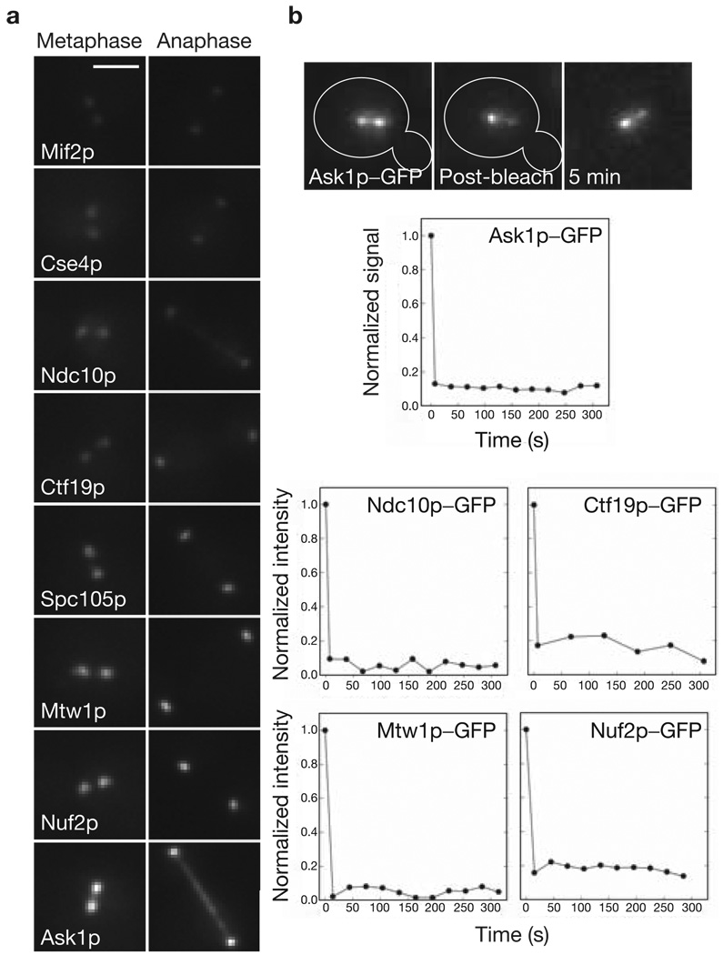
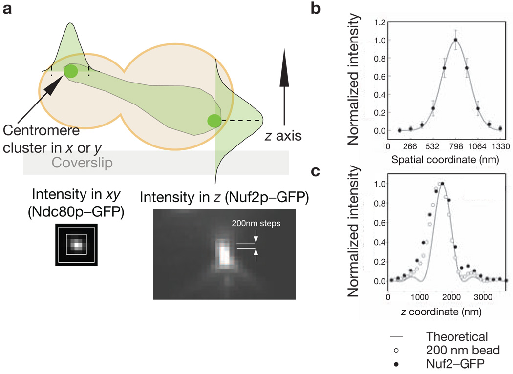
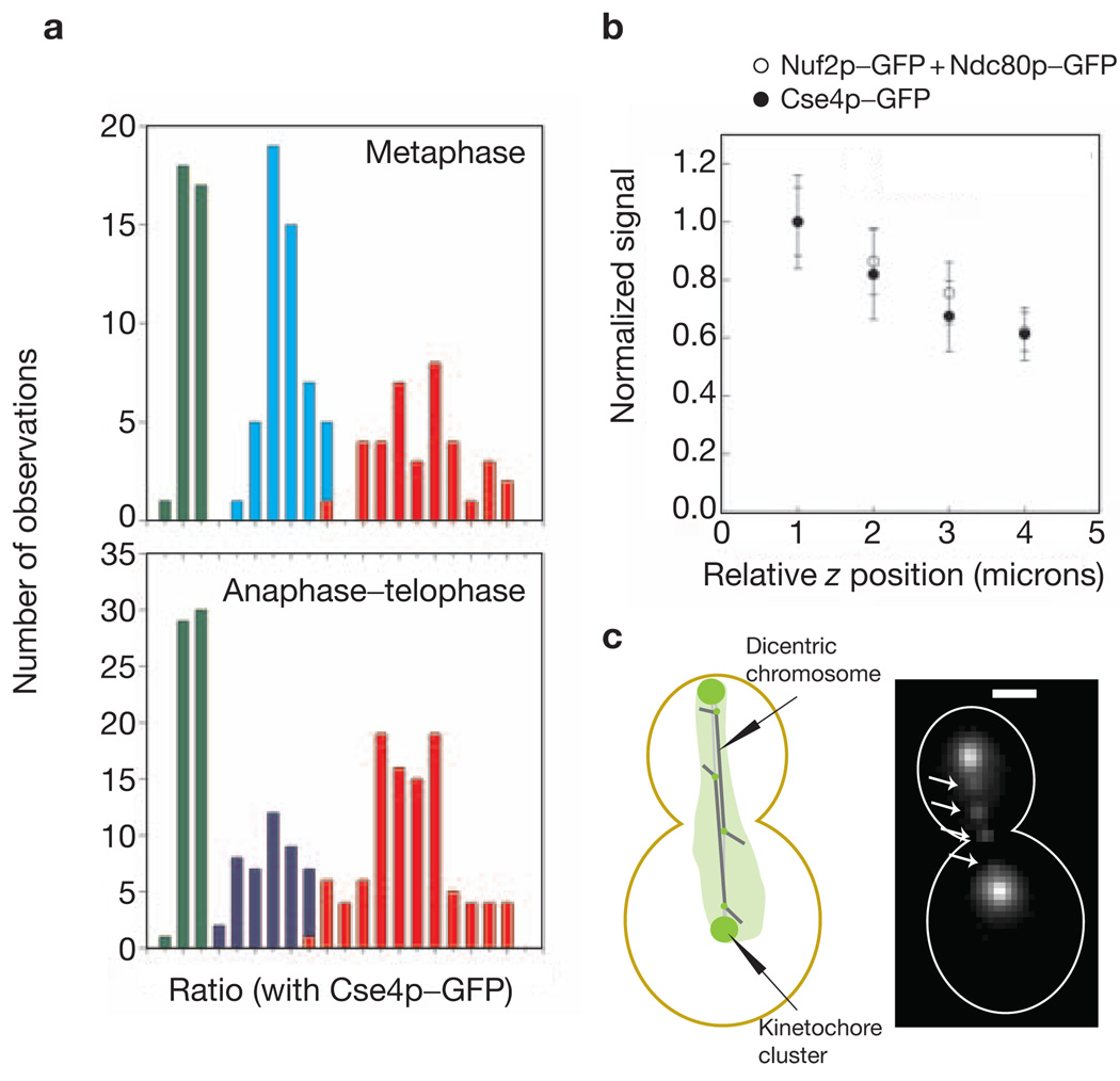
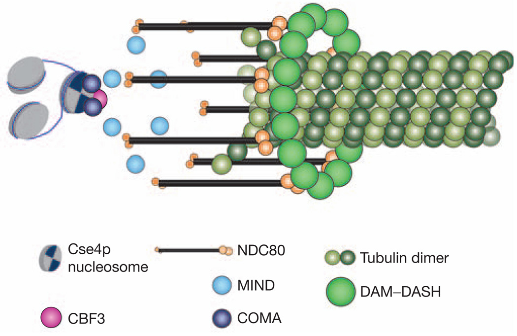
Kinetochore attachment to spindle microtubule plus-ends is necessary for accurate chromosome segregation during cell division in all eukaryotes. The centromeric DNA of each chromosome is linked to microtubule plus-ends by eight structural-protein complexes. Knowing the copy number of each of these complexes at one kinetochore-microtubule attachment site is necessary to understand the molecular architecture of the complex, and to elucidate the mechanisms underlying kinetochore function. We have counted, with molecular accuracy, the number of structural protein complexes in a single kinetochore-microtubule attachment using quantitative fluorescence microscopy of GFP-tagged kinetochore proteins in the budding yeast Saccharomyces cerevisiae. We find that relative to the two Cse4p molecules in the centromeric histone, the copy number ranges from one or two for inner kinetochore proteins such as Mif2p, to 16 for the DAM-DASH complex at the kinetochore-microtubule interface. These counts allow us to visualize the overall arrangement of a kinetochore-microtubule attachment. As most of the budding yeast kinetochore proteins have homologues in higher eukaryotes, including humans, this molecular arrangement is likely to be replicated in more complex kinetochores that have multiple microtubule attachments.


Figures

Figure 1
Figure 1
Localization and turnover of kinetochore proteins in metaphase and in anaphase–telophase. (a) Localization of representative GFP-tagged kinetochore proteins during metaphase and anaphase–telophase. At metaphase, the sister kinetochores become aligned on either side of the spindle equator into two distinct clusters each containing 16 kinetochores. Note that both CBF3 and the DAM–DASH complex also localize to the spindle in anaphase. The scale bar represents 2 µm. (b) Pre- and post-photobleaching images of a metaphase cell expressing Ask1p–GFP. Signal recovery (shown in the relative intensity versus time graphs) is undetectable for at least 300 s. Recovery is similarly low for representative proteins from four other complexes.
Figure 2
Figure 2
Characterization of the intensity distribution of a kinetochore cluster. (a) A schematic representation of a budding yeast cell expressing a GFP-tagged kinetochore protein in anaphase–telophase is shown. Signal was measured by integrating the signal intensity in the xy direction (shown for Ndc80p–GFP) in the plane that contains the maximum intensity pixel along the z axis (shown for Nuf2p–GFP). (b) Fitting a Gaussian function to the anaphase–telophase intensity distribution in the xy plane for Nuf2p–GFP yields σ = 159 nm (s.d. for the Gaussian curve). Similar measurement for metaphase xy intensity distribution yields σ = 189 nm. (c) The intensity distribution along the z axis for a Nuf2p-GFP cluster and a 200 nm green fluorescent bead. The solid line represents the theoretical intensity distribution along the z axis.
Figure 3
Figure 3
Linearity and sensitivity of the measurement technique. (a) A frequency histogram for signal measurements in metaphase and anaphase–telophase for Cse4p–GFP (green), Nuf2p–GFP (cyan), Ndc80p–GFP (blue) and Nuf2p–GFP + Ndc80p–GFP (red). The proportional increase in the fluorescence intensity for Nuf2p–GFP + Ndc80p–GFP also demonstrates that the proximity of fluorophores does not detectably affect their fluorescence. (b) Normalized signal plotted as a function of the relative z coordinate of the kinetochore clusters of Cse4p–GFP and Nuf2p–GFP + Ndc80p–GFP. The error bars represent the s.d. of the mean signal value in each bin. (c) Four lagging kinetochores (arrows, Nuf2p–GFP) on the two dicentric chromosomes in a mid-anaphase cell. The chromatin between the two centromeres is 40 kb long. The scale bar represents 2 µm.
Figure 4
Figure 4
A schematic representation of the possible architecture of a kinetochore–microtubule attachment site in metaphase. This structure is based on the protein counts reported here, and on the in vitro structures of the DAM–DASH and the NDC80 complex. It depicts only five essential kinetochore protein complexes.

References

    1. Meluh PB, Yang P, Glowczewski L, Koshland D, Smith MM. Cse4p is a component of the core centromere of Saccharomyces cerevisiae. Cell. 1998;94:607–613. - PubMed
    1. Meluh PB, Koshland D. Evidence that the MIF2 gene of Saccharomyces cerevisiae encodes a centromere protein with homology to the mammalian centromere protein CENP-C. Mol. Biol. Cell. 1995;6:793–807. - PMC - PubMed
    1. Espelin CW, Kaplan KB, Sorger PK. Probing the architecture of a simple kinetochore using DNA–protein crosslinking. J. Cell Biol. 1997;139:1383–1396. - PMC - PubMed
    1. Ortiz J, Stemmann O, Rank S, Lechner J. A putative protein complex consisting of Ctf19, Mcm21, and Okp1 represents a missing link in the budding yeast kinetochore. Genes Dev. 1999;13:1140–1155. - PMC - PubMed
    1. Nekrasov VS, Smith MA, Peak-Chew S, Kilmartin JV. Interactions between centromere complexes in Saccharomyces cerevisiae. Mol. Biol. Cell. 2003;14:4931–4946. - PMC - PubMed

Publication types

MeSH terms

Substances