Comparative Study
. 2006 Jun;59(6):952-62.
doi: 10.1002/ana.20873.

Frontotemporal dementia: clinicopathological correlations

Affiliations
Comparative Study

Frontotemporal dementia: clinicopathological correlations

Mark S Forman et al. Ann Neurol. 2006 Jun.

Abstract

Objective: Frontotemporal lobar degeneration (FTLD) is characterized by impairments in social, behavioral, and/or language function, but postmortem studies indicate that multiple neuropathological entities lead to FTLD. This study assessed whether specific clinical features predict the underlying pathology.

Methods: A clinicopathological correlation was performed on 90 consecutive patients with a pathological diagnosis of frontotemporal dementia and was compared with an additional 24 cases accrued during the same time period with a clinical diagnosis of FTLD, but with pathology not typically associated with frontotemporal dementia.

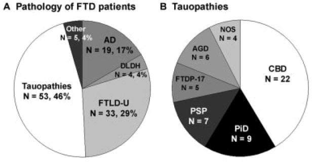
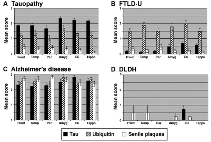
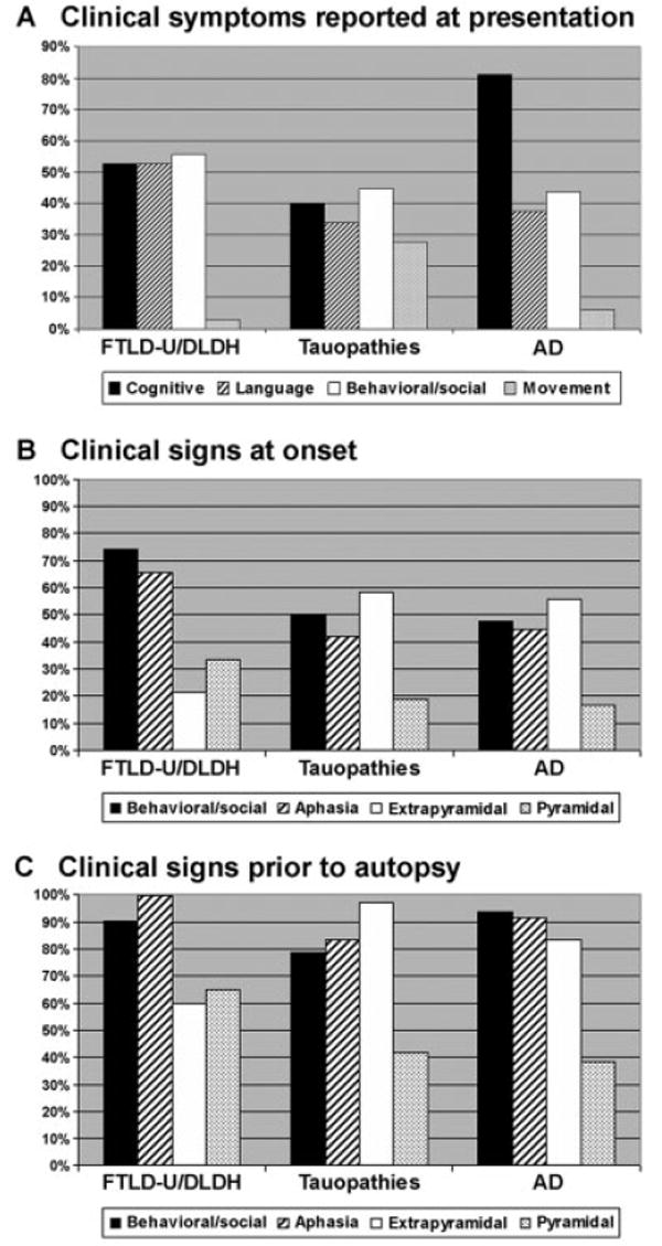
Results: Postmortem examination showed multiple pathologies including tauopathies (46%), FTLD with ubiquitin-positive inclusions (29%), and Alzheimer's disease (17%). The pathological groups manifested some distinct demographic, clinical, and neuropsychological features, although these attributes showed only a statistical association with the underlying pathology. FTLD with ubiquitin-positive inclusions was more likely to present with both social and language dysfunction, and motor neuron disease was more likely to emerge in these patients. Tauopathies were more commonly associated with an extrapyramidal disorder. Alzheimer's disease was associated with relatively greater deficits in memory and executive function.

Interpretation: Clinical and neuropsychological features contribute to delineating the spectrum of pathology underlying a patient diagnosed with FTLD, but biomarkers are needed that, together with the clinical phenotype, can predict the underlying neuropathology.


Figures

Fig 1
Fig 1
Algorithm for the neuropathological diagnosis of patients with frontotemporal lobar degeneration (FTLD). This algorithm represents an oversimplification of the scheme used to diagnose FTLD patients and does not reflect the complexity of the cases or the commonly observed occurrence of multiple pathologies. 3R-tau, 4R-tau, and 3R/4R-tau refer to the specific subsets of insoluble tau isoforms observed biochemically and characterized by either three or four microtubule binding repeats. AD = Alzheimer’ disease; AGD = argyrophilic grain disease; CBD = corticobasal degeneration; DLDH = dementia lacking distinctive histopathology; FTDP-17 = frontotemporal dementia with parkinsonism linked to chromosome 17; FTLD-U = frontotemporal lobar degeneration with ubiquitin-positive inclusions; LBVAD = Lewy body variant of Alzheimer’s disease; MND = motor neuron disease; NFT = neurofibrillary tangle; NIFID = neuronal intermediate filament inclusion dementia; PiD 3 Pick’s disease; PSP = progressive supranuclear palsy; TPSD = tangle-predominant senile dementia.
Fig 2
Fig 2
Neuropathology of frontotemporal dementia (FTD). (A) Primary neuropathological diagnosis of patients who presented clinically with frontotemporal lobar degeneration (FTLD). The number of patients and percentage of total are indicated. The “other” category includes patients with Lewy body disease (n = 2), Creutzfeldt–Jakob disease (n = 1), cerebrovascular disease (n = 1), and normal adult brain (n = 1). Additional, less robust pathology was present in 20.4% of patients and consisted of either a second, less robust, neurodegenerative disorder present in 13.8% of patients or cerebrovascular disease present in 6.6% of patients. (B) Neuropathological subtypes of tauopathies. The number of patients in each category is indicated. AD = Alzheimer’ disease; AGD = argyrophilic grain disease; CBD = corticobasal degeneration; DLDH = dementia lacking distinctive histopathology; FTDP-17 = frontotemporal dementia with parkinsonism linked to chromosome 17; FTLD-U = frontotemporal lobar degeneration with ubiquitin-positive inclusions; NOS = not otherwise specified; PiD = Pick’s disease; PSP = progressive supranuclear palsy.
Fig 3
Fig 3
Topographical distribution of pathological inclusions in frontotemporal dementia (FTD) subgroups. Semiquantitative distribution of tau (black bars), ubiquitin (striped bars), and senile plaque (white bars) inclusion pathology in FTD subgroups. The tau pathology in the Alzheimer’s disease (AD) patients was largely Thioflavine S–positive, whereas in the tauopathies, the pathology was largely Thioflavine S–negative. This is reflected in the Braak stage for each of the subgroups: (A) tau = 1.7 ± 1.1; (B) frontotemporal lobar degeneration with ubiquitin-positive inclusions (FTLD-U) = 0.8 ± 0.7; (C) AD = 5.8 ± 0.8; (D) dementia lacking distinctive histopathology (DLDH) = 0.8 ± 0.5. Amyg = amygdala; EC = entorhinal cortex; Front = middle frontal gyrus; Hippo = hippocampus, CA1/subiculum; Par = inferior parietal lobule; Temp = superior temporal gyrus.
Fig 4
Fig 4
Clinical symptoms and signs distinguish pathological subgroups of frontotemporal dementia (FTD). (A) The percentage of patients and/or informants reporting the presence of the clinical features indicated. When stratified by pathological diagnosis, the presenting symptoms showed patterns that distinguish the various pathological subgroups. Behavioral/social (white bars) = disinhibition, change in personality, apathy, decreased motivation, and psychiatric illness diagnosed later in life near the onset of symptoms; language disorders (striped bars) = naming or word-finding difficulty, decline in speech or grammar; cognitive deficits (black bars) = memory loss, decline in activities of daily living, attention deficits, executive dysfunction, or visuospatial complaints; movement problems (gray bars) = abnormal gait, falling, rigidity, apraxia, or dysarthria. (B, C) The percentage of patients diagnosed at first (B) and last (C) clinical examination in the major pathological subgroups with language (striped bars), behavioral/social (black bars), extrapyramidal (white bars), and/or pyramidal dysfunction (gray bars) is indicated. Although the pattern of clinical signs did not significantly distinguish the pathological subgroups, the presence of both social dysfunction and aphasia at first examination was specific for frontotemporal lobar degeneration with ubiquitin-positive inclusions/dementia lacking distinctive histopathology (FTLD-U/DLDH) compared with Alzheimer’s disease (AD) and tauopathy groups. Extrapyramidal symptoms were more common in the tauopathy subgroups (58%) relative to the FTLD-U/DLDH subgroups (21%), and pyramidal symptoms were frequently identified in FTLD-U/ DLDH patients, although this difference did not reach statistical significance. With disease progression, the incidence of social dysfunction, aphasia, extrapyramidal signs, and pyramidal signs increased in all subgroups to a common status of global impairment in all pathology subgroups.

References

    1. Neary D, Snowden JS, Gustafson L, et al. Frontotemporal lobar degeneration: a consensus on clinical diagnostic criteria. Neurology. 1998;51:1546–1554. - PubMed
    1. Grossman M. Frontotemporal dementia: a review. J Int Neuropsychol Soc. 2002;8:566–583. - PubMed
    1. McKhann GM, Albert MS, Grossman M, et al. Clinical and pathological diagnosis of frontotemporal dementia: report of the Work Group on Frontotemporal Dementia and Pick’s Disease. Arch Neurol. 2001;58:1803–1809. - PubMed
    1. Grossman M, Ash S. Primary progressive aphasia: a review. Neurocase. 2004;10:3–18. - PubMed
    1. Hodges JR, Patterson K, Oxbury S, et al. Semantic dementia. Progressive fluent aphasia with temporal lobe atrophy. Brain. 1992;115(pt 6):1783–1806. - PubMed

Publication types

MeSH terms