

Splenic vasculopathy in portal hypertension patients
- PMID: 16718761
- PMCID: PMC4130983
- DOI: 10.3748/wjg.v12.i17.2737
Splenic vasculopathy in portal hypertension patients
Abstract
Aim: To investigate the interaction between portal hypertension, splanchnic hyperdynamic circulation and splanchnic vasculopathy by observing splenic arterial and venous pathological changes and the ro1e of extra-cellular matrix in the pathogenesis of portal hypertensive vasculopathy by measuring the expression of type I and type III procollagen mRNA in splenic venous walls of portal hypertensive patients.
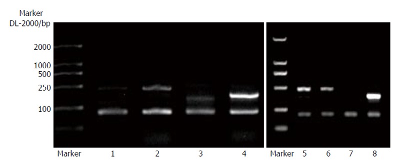
Methods: Morphological changes of splenic arteries and veins taken from portal hypertensive patients (n=20) and normal controls (n=10) were observed under optical and electron microscope. Total RNA was extracted and the expression of type I and type III procollagen mRNA in splenic venous walls of portal hypertensive patients (n=20) was semi-quantitatively detected using reverse transcription-polymerase chain reaction (RT-PCR).
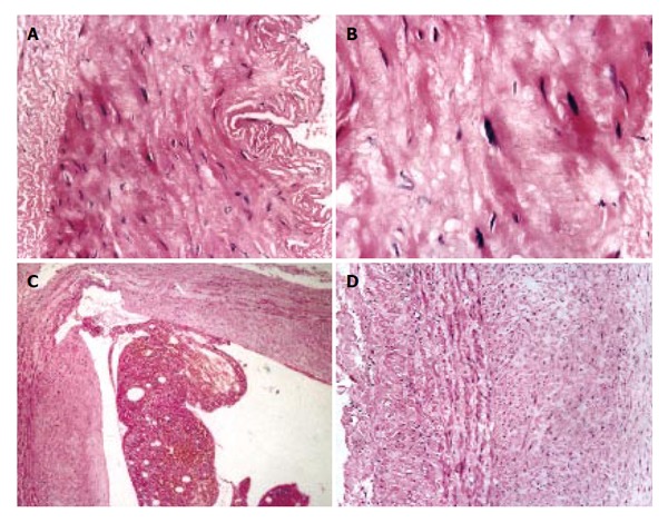
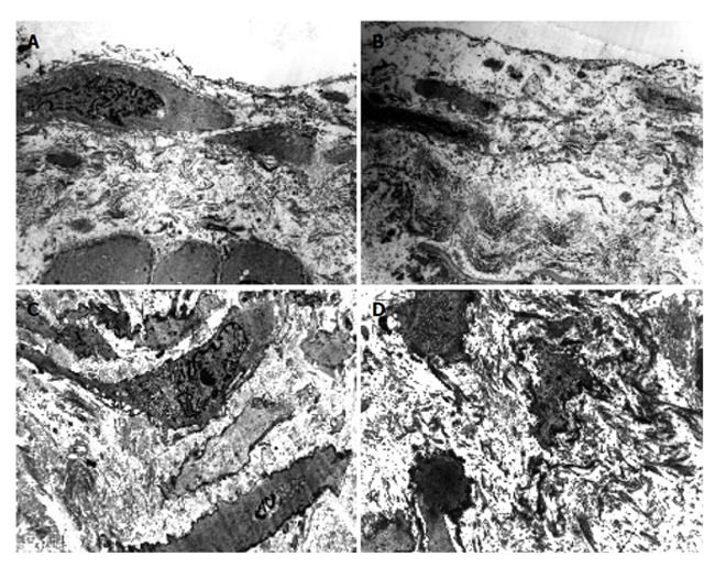
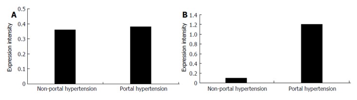
Results: Under optical microscope, splenic arterial intima was destroyed and internal elastic membrane and medial elastic fibers of the splenic arterial walls were degenerated and broken. Splenic venous intima became remarkably thick. Endothelia1 cells were not intact with formation of mural thrombus. The tunica media became thickened significantly due to hypertrophy of smooth muscles. Fibers and connective tissues were increased obviously. Under electron microscope, smooth muscle cells of the splenic arteries were degenerated and necrotized. Phenotypes of smooth muscle cells changed from constrictive into synthetic type. Red blood cells and platelets accumulated around the damaged endothelial cells. Synthetic smooth muscle cells were predominant in splenic veins and their cytoplasma had plentiful rough endoplasmic reticulum ribosomes and Golgi bodies. Along the vascular wall, a lot of collagen fibers were deposited, the intima was damaged and blood components accumulated. There was no significant difference in the expression of type I procollagen mRNA in splenic venous wall between the patients with portal hypertension and those without portal hypertension (P>0.05), but the expression of type III procoagen mRNA was significantly stronger in the patients with portal hypertension than in those without portal hypertension (P<0.01).
Conclusion: Type III procollagen and collagen might be important extra-cellular matrix resulting in neointimal formation and vascular remodeling in the pathogenesis of portal hypertensive vasculopathy. The pathological changes in splenic arteries and veins exist in portal hypertension patients. There might be an interaction between portal hypertension, splanchnic hyperdynamic circulation and splanchnic vasculopathy.
Figures
References
-
- Yang Z, Ren D, Li D, Qiu F. [Portal hypertensive vasculopathy of splenic artery] Zhonghua Waike Zazhi. 1999;37:412–414. - PubMed
-
- Yang Z, Zhang L, Li D, Qiu F. Pathological morphology alteration of the splanchnic vascular wall in portal hypertensive patients. Chin Med J (Engl) 2002;115:559–562. - PubMed
-
- Lincoln TM, Dey N, Sellak H. Invited review: cGMP-dependent protein kinase signaling mechanisms in smooth muscle: from the regulation of tone to gene expression. J Appl Physiol. 2001;91:1421–1430. - PubMed
-
- Hayashi K, Takahashi M, Nishida W, Yoshida K, Ohkawa Y, Kitabatake A, Aoki J, Arai H, Sobue K. Phenotypic modulation of vascular smooth muscle cells induced by unsaturated lysophosphatidic acids. Circ Res. 2001;89:251–258. - PubMed
-
- Jiang B, Xu S, Brecher P, Cohen RA. Growth factors enhance interleukin-1 beta-induced persistent activation of nuclear factor-kappa B in rat vascular smooth muscle cells. Arterioscler Thromb Vasc Biol. 2002;22:1811–1816. - PubMed
Publication types
MeSH terms
Substances
LinkOut - more resources
Full Text Sources
