

Review
doi: 10.3748/wjg.v12.i18.2858.
Morphology and motor function of the gastrointestinal tract examined with endosonography
Affiliations
- PMID: 16718809
- PMCID: PMC4087801
- DOI: 10.3748/wjg.v12.i18.2858
Item in Clipboard
Review
Morphology and motor function of the gastrointestinal tract examined with endosonography
World J Gastroenterol.
.
Abstract
Endosonography is a useful tool for studying the morphology and motor function of the gastrointestinal tract. Intraluminal ultrasonography is the common denomination of ultrasound examinations using intracorporal transducers which are inserted into the GI tract. Thus, the visceral wall and adjacent structures can be imaged in detail. This review describes the usefulness of endosonography in gastroenterology, in particular with respect to studies of the biomechanical and motor function of the gastrointestinal tract. New techniques such as 3-D EUS, elastography and strain rate imaging are discussed.
Figures
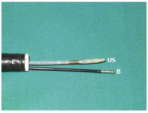
An endosco-pe with two channels. A miniature ultrasound transducer (US) and a biopsy forceps (B) are inserted through the channels.
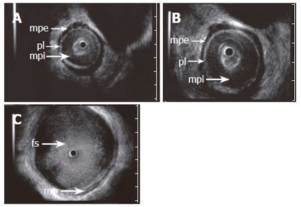
ES images (A and B) obtained with a 15 MHz miniprobe in a patient with achalasia showing the longitudinal, outer layer of the proper muscle (mpe) separated from a thickened circular, inner layer (mpi) by an echogenic layer corresponding to the localization of the plexus myentericus (pl). C: shows a dilated esophageal lumen with food content (fs). A normal aspect of the proper muscle is seen.
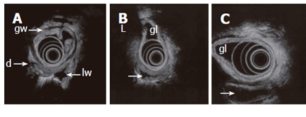
Endosonography with an echoendoscope (12 MHz frequency is used) placed in the gastric lumen (gl) showing the layers of the gastric wall (gw) and GI loops outside the stomach. Lumen is partly collapsed (d), partly waterfilled (arrows). Motility and contractions of native GI loops may be studied. High frequency miniprobes are less suitable for this purpose due to higher frequencies and small transducer diameter.
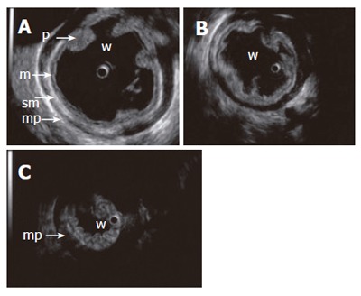
Contraction sequence (A, B, C) demonstrated with a 15 MHz miniprobe in a water-filled (w) stomach. Gastric fold (p), mucosa (m), submucosa (sm), proper muscle (mp).

A normal pancreas imaged from the stomach with a 6 MHz echoendoscope. The compressed posterior gastric wall is closely related to the surface of the pancreas. The pancreatic duct is seen (arrow) with minor (A) and slightly increased transducer pressure (B). The non-compressed gastric wall with folds (gw) is seen on the opposite side of the transducer. Pancreatic tail (pt), splenic vein (sv).
References
-
- Tio TL. Endosonography in gastroenterology. Berlin: Springer-Verlag; 1988.
-
- Rösch T, Classen M. Gastroenterologic endosonography. Stuttgart: Georg Thieme Verlag; 1992.
-
- Van Dam J, Sivak MV, eds . Gastrointestinal endosonography. Philadelphia: W.B. Saunders Company; 1999.
-
- Giovannini M, Bories E, Pesenti C, Monges G, Moutardier V, Delpero J. Elastography and contrast-enhanced (Sonovue) color. Doppler pancreatic endoscopic ultrasound to characterize pancreatic mass. Results in 25 patients. Endoscopy. 2005;37 Suppl I:A68.
-
- Matre K, Odegaard S, Hausken T. Endoscopic ultrasound Doppler probes for velocity measurements in vessels in the upper gastrointestinal tract using a multifrequency pulsed Doppler meter. Endoscopy. 1990;22:268–270. - PubMed
Publication types
MeSH terms
LinkOut - more resources
Full Text Sources
