. 2006 Jun;4(6):e186.
doi: 10.1371/journal.pbio.0040186.

Control of phage Bxb1 excision by a novel recombination directionality factor

Affiliations

Control of phage Bxb1 excision by a novel recombination directionality factor

Pallavi Ghosh et al. PLoS Biol. 2006 Jun.

Abstract

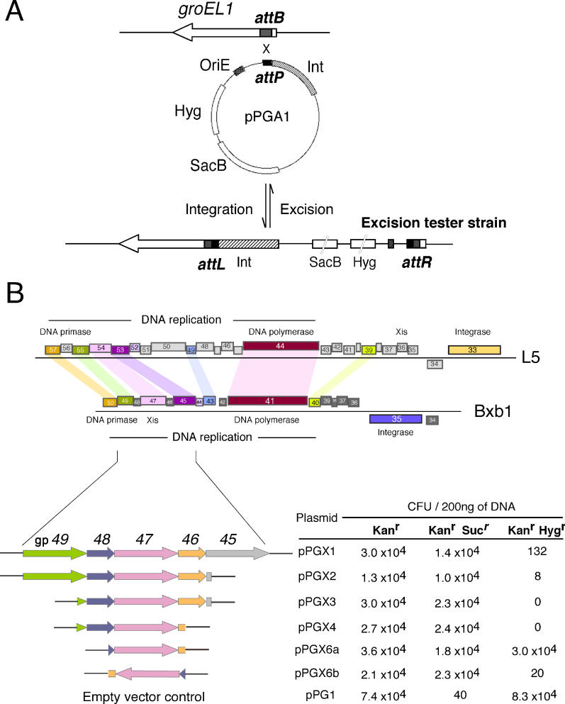
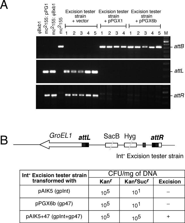
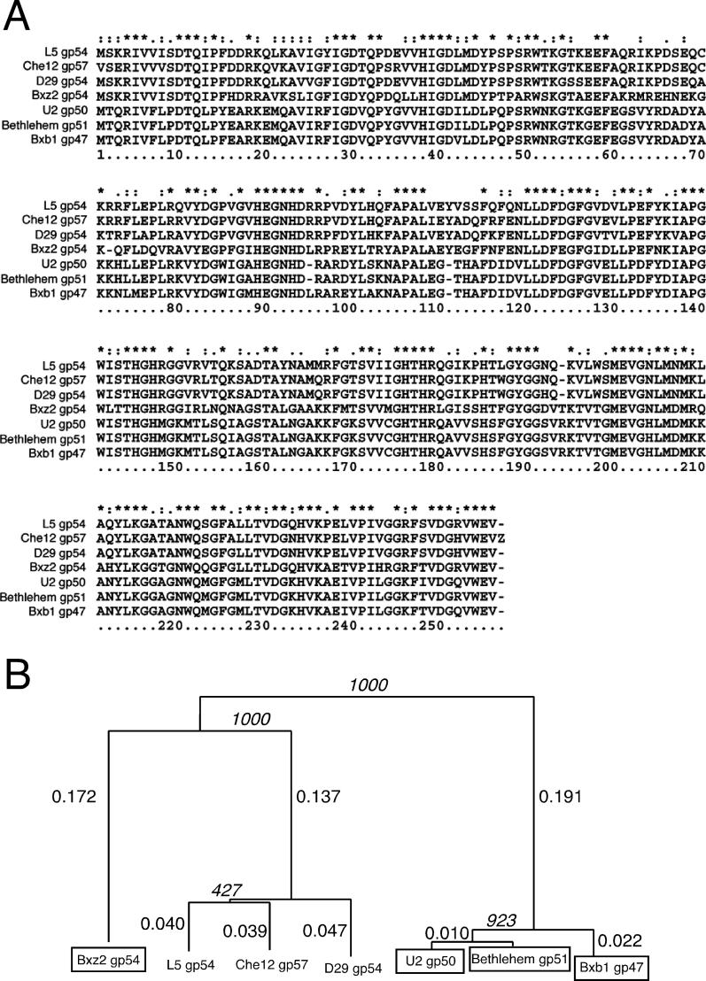
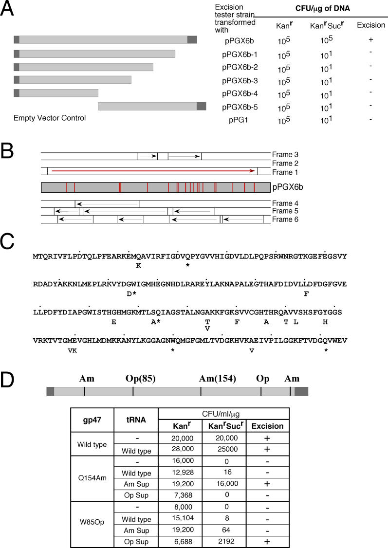
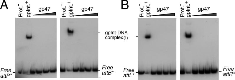
Mycobacteriophage Bxb1 integrates its DNA at the attB site of the Mycobacterium smegmatis genome using the viral attP site and a phage-encoded integrase generating the recombinant junctions attL and attR. The Bxb1 integrase is a member of the serine recombinase family of site-specific recombination proteins and utilizes small (<50 base pair) substrates for recombination, promoting strand exchange without the necessity for complex higher order macromolecular architectures. To elucidate the regulatory mechanism for the integration and excision reactions, we have identified a Bxb1-encoded recombination directionality factor (RDF), the product of gene 47. Bxb1 gp47 is an unusual RDF in that it is relatively large (approximately 28 kDa), unrelated to all other RDFs, and presumably performs dual functions since it is well conserved in mycobacteriophages that utilize unrelated integration systems. Furthermore, unlike other RDFs, Bxb1 gp47 does not bind DNA and functions solely through direct interaction with integrase-DNA complexes. The nature and consequences of this interaction depend on the specific DNA substrate to which integrase is bound, generating electrophoretically stable tertiary complexes with either attB or attP that are unable to undergo integrative recombination, and weakly bound, electrophoretically unstable complexes with either attL or attR that gain full potential for excisive recombination.


Figures

Figure 1
Figure 1. Identification of the Bxb1 RDF
(A) The hygromycin-resistance cassette, sucrose-sensitivity cassette (sacB), and a portion of the Bxb1 DNA containing attP and int were cloned into an integrative plasmid containing the plasmid ColE1 origin of E. coli (OriE) , and the resultant plasmid pPGA1 was transformed into M. smegmatis mc 2155. Expression of int drives integration of the plasmid into the host attB site leading to the formation of attL and attR sites flanking the hyg and sacB cassettes. The resultant strain, designated as the excision tester strain, is resistant to hygromycin and sensitive to the presence of sucrose. An excisive recombination event between attL and attR results in the removal (and subsequent loss) of the intervening DNA containing int, hyg, and sacB; the strain consequently becomes hyg S and suc R and can thereby be monitored by the appearance of colonies in the presence of sucrose. (B) A segment of the Bxb1 genome containing genes involved in integration and DNA replication as well as the corresponding portion of the phage L5 genome is shown in the upper part; related genes are colored accordingly. The lower part of the figure shows the region of Bxb1 DNA present in plasmids exhibiting excision activity. Following isolation of plasmid pPGX1—which is active in promoting sucrose resistance in the excision tester strain shown in (A)—plasmid derivatives containing deletions from both ends of pPGX1 were constructed, introduced into the excision tester strain, and scored for the appearance of suc R colonies. CFU, colony-forming units
Figure 2
Figure 2. Confirmation of Site-Specific Recombination and Bxb1 Integrase Dependence
(A) DNA from five sucrose-resistant transformants of the excision tester strain transformed with either pPG1 (empty vector), pPGX1, or pPGX6b was examined by PCR for the presence of attB, attL, and attR. Transformation with pPGX1 and pPGX6b leads to the presence of a product amplified with attB-specific primers; no product is observed using primers that amplify attL and attR. DNA from a Bxb1 lysogen and from clones transformed with the empty vector that were used as controls show a product corresponding to attL and attR, whereas DNA from wild-type mc 2155 shows the presence of a product corresponding to attB. (B) An int excision tester strain was created (as in Figure 1A) using transient expression of gpInt. The int tester strain was then transformed with pAIK5 (gpInt alone), pX6b (gp47 alone), or pAIK5+47 (gpInt + gp47), and the frequency of suc R colonies (and therefore excision) determined. CFU, colony-forming units
Figure 3
Figure 3. Sequence Alignment of Bxb1 gp47 and Its Homologs
(A) A BLAST search identified proteins of mycobacteriophages U2 (gp50), Bethlehem (gp51), L5 (gp54), D29 (gp54), Che12 (gp57), and Bxz2 (gp54) with similarity to Bxb1 gp47. Multiple sequence alignment of these proteins was performed using ClustalW (identical residues indicated by an asterisk [*]) and the phylogenetic relationship (B) represented using NJPLOT; branch lengths and bootstrap (italicized) values are indicated. The proteins encoded by phages that encode a serine integrase (Bxz2, U2, Bethlehem, and Bxb1) are boxed; all others (L5, D29, and Che12) encode a tyrosine integrase.
Figure 4
Figure 4. Confirmation of Bxb1 gp47 as the Phage RDF
(A) The solid bar shows the portion of Bxb1 DNA from 35,976–36,811 bp contained within pPGX6b with the regions flanking gene 47 in dark grey. Truncation derivatives of pPGX6b were constructed as shown and tested for excision activity as in Figure 1. The number of sucR colonies obtained upon transformation of the excision tester strain with each of the derivatives is shown. (B) Plasmid pPGX6b was randomly mutagenized by passage through a mutator strain, and excision defective mutants were selected ( Figure 1). The positions of 20 excision-defective mutants are shown as solid vertical lines (red), all of which lie within gene 47. Arrows indicate ORFs in all six reading frames of pPGX6b, with Bxb1 gene 47 shown in red. (C) The amino acid changes in Bxb1 gp47 corresponding to each of the 15 base changes in pPGX6b are shown; the five nonsense substitutions are marked with an asterisk (*). (D) The locations of five nonsense mutations inactive in excision are shown using solid vertical lines. An excision tester strain previously transformed with plasmids expressing gp47 (wild type) or either of the two nonsense mutations (Q154Am or W85Op) was transformed with plasmids expressing nonsense suppressors generated by modification of tRNAs encoded by mycobacteriophage L5 [ 41] (as indicated) and scored for the appearance of sucR colonies; the numbers of sucR colonies are shown. CFU, colony-forming units
Figure 5
Figure 5. In Vitro Excisive Recombination Using gp47
(A) E. coli BL21(DE3)pLysS transformed with pET28a and pPGgp47 were grown to an A 600 of 0.6 at 30 °C and induced for an additional 4 h at 22 °C with 0.6 mM IPTG. The cells were lysed in lysis buffer (see Materials and Methods) and partially purified by passage through a Ni-NTA column followed by elution with 150 mM imidazole; I, S, Ft, W, and E represent the insoluble fraction, soluble fraction, flow-through from the Ni-NTA column, washes with the indicated concentration of imidazole, and 150 mM elution from the Ni-NTA column, respectively. The induced cells of pPGgp47 show the presence of an approximately 32-kDa protein (as indicated) that is absent from the pET28a expression lanes and is abundant in the insoluble fraction. Molecular weight markers are shown in lane M and their corresponding sizes indicated. (B) Integrative recombination was performed as described previously using a supercoiled attP substrate, a linear 50-bp attB DNA, and increasing concentrations of gpInt [ 34]: panel a, in the absence of any additional protein; panel b, in the presence of partially purified gp47; panel c, with addition of a control extract. Lanes 1–5 contain 0.36, 0.18, 0.09, 0.045, and 0.0225 μM of gpInt respectively. Panels b and c contain 1.78 μM of gp47 and an equivalent amount of the control extract respectively, in addition to gpInt. The positions of the supercoiled substrate and the linear recombinant product are indicated. The small (50 bp) linear attB substrate migrates fast and is not shown. (C) Excisive recombination was carried out in recombination buffer (see Materials and Methods) using a 367-bp attL in a supercoiled plasmid and a 377-bp linear attR partner DNA. Lanes 1–5 of panel a contain increasing concentrations of gp47 (0.89, 1.78, 2.67, 3.56, and 5.34 μM), lanes 7–11 of panel b contain an equivalent amount of the control protein. Control reactions lacking either the partner attR DNA (lane12), gp47 (lane 13), or gpInt (lane14) are shown in panel c. The positions of the supercoiled attL substrate and the linear recombinant product are indicated. (D) Excisive recombination reactions with varying sizes of linear DNA substrates show that only small substrate sites are required. Panel a shows recombination between a supercoiled plasmid containing a 367-bp attL and varying sizes of linear attR partner DNA as indicated. Panel b shows recombination between a supercoiled plasmid containing a 377-bp attR and varying sizes of linear attL partner DNAs. The positions of supercoiled substrates and linear recombinant products are indicated.
Figure 6
Figure 6. . DNA-Binding Properties of Bxb1 gp47
(A and B) The ability of gp47 to bind attP, attB, attL, and attR was determined using native gel electrophoresis. Binding of gp47 or gpInt to attP and attB (A) or attL and attR (B) was performed with either gpInt (0.072 μM), or increasing concentrations of gp47 (0.45, 0.89, 1.78, and 3.56 μM).
Figure 7
Figure 7. Substrate-Dependent Interaction of Bxb1 gpInt and gp47
(A and B) Binding of gpInt and gp47 to attP/ attB/ attL/ attR was monitored by native gel electrophoresis. Radiolabeled DNA fragments (˜300 bp) were incubated with either gpInt (0.072 μM) alone or gpInt with increasing concentrations of gp47 (0.45, 0.89, 1.78, and 3.56 μM). The positions of DNA–gpInt complexes (cmplx I) as well as tertiary complexes containing DNA, gpInt, and gp47 (cmplx II) are shown. (C and D) The presence of gp47 in the tertiary complexes shown in (A) was determined by the ability of α-His antibodies to supershift complexes observed by native gel electrophoresis. α-His antibodies were either added to reactions containing DNA, gpInt, and gp47 (indicated as lane 1), or first preincubated with gp47 for 30 min and then added to reactions containing DNA and gpInt (lane 2). The protein–DNA complexes were separated from free DNA on a 5% native PAGE. The positions of the tertiary complexes of gp47, gpInt, and DNA as well as the antibody supershifted complexes are indicated. (E and F) Bxb1 gp47 is required for trapping a synaptic complex in excision. A suicide substrate version of attL DNA (5′ radiolabeled at both ends) was used that has a nick on the top strand positioned four bases to the 5′ side of the scissile bond. Bxb1 gpInt (72 nM) binds normally to this substrate to form Complex I (cmplx I), but when attR partner DNA (200 bp) and gp47 (3.56 μM) is added, no recombinant products are released. Instead, a prominent slow-moving complex is observed that absolutely requires Bxb1 gp47 for its formation. We have identified this as a synaptic complex using 2D-PAGE (F). In brief, a vertical gel slice was removed from the last lane in panel E, incubated with proteinase K and SDS, and then electrophoresed through a second dimension. Approximately 50% of the radiolabeled DNAs in this complex correspond to attP recombinant product and 50% correspond to a cleaved half-site. The bottom of the gel slice containing unbound attL DNA was removed prior to the second dimension of electrophoresis. Further details on the characterization of these suicide substrates will be described in future publications.

Comment in

References

    1. Echols H. Lysogeny: Viral repression and site-specific recombination. Annu Rev Biochem. 1971;40:827–854. - PubMed
    1. Nash HA. Integration and excision of bacteriophage lambda: The mechanism of conservation site specific recombination. Annu Rev Genet. 1981;15:143–167. - PubMed
    1. Lewis JA, Hatfull GF. Control of directionality in integrase-mediated recombination: Examination of recombination directionality factors (RDFs) including Xis and Cox proteins. Nucleic Acids Res. 2001;29:2205–2216. - PMC - PubMed
    1. Weisberg RA, Landy A. Site specific recombination in phage lambda. In: Hendrix RW, Roberts JW, Stahl FW, Weisberg RA, editors. Lambda II. Cold Spring Harbor (New York): Cold Spring Harbor Laboratory; 1983. pp. 211–250.
    1. Landy A. Dynamic, structural, and regulatory aspects of lambda site-specific recombination. Annu Rev Biochem. 1989;58:913–949. - PubMed

Publication types

MeSH terms