. 2006 May 23:6:22.
doi: 10.1186/1471-213X-6-22.

Expression of carbonic anhydrases IX and XII during mouse embryonic development

Affiliations

Expression of carbonic anhydrases IX and XII during mouse embryonic development

Heini Kallio et al. BMC Dev Biol. .

Abstract

Background: Of the thirteen active carbonic anhydrase (CA) isozymes, CA IX and XII have been linked to carcinogenesis. It has been suggested that these membrane-bound CAs participate in cancer cell invasion, which is facilitated by an acidic tumor cell environment. Since active cell migration is a characteristic feature of embryonic development, we set out to explore whether these isozymes are expressed in mouse embryos of different ages. The studies were focused on organogenesis stage.

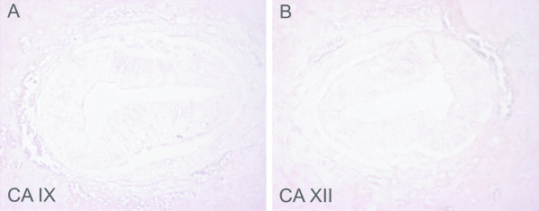
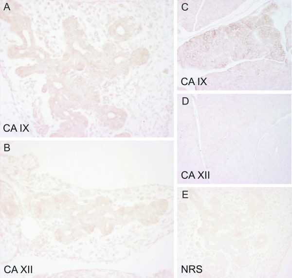
Results: Immunohistochemistry demonstrated that both CA IX and XII are present in several tissues of the developing mouse embryo during organogenesis. Staining for CA IX revealed a relatively wide distribution pattern with moderate signals in the brain, lung, pancreas and liver and weak signals in the kidney and stomach. The expression pattern of CA XII in the embryonic tissues was also relatively broad, although the intensity of immunostaining was weak in most tissues. The CA XII-positive tissues included the brain, where the most prominent staining was seen in the choroid plexus, and the stomach, pancreas, liver and kidney.

Conclusion: Membrane-bound CA isozymes IX and XII are expressed in various tissues during mouse organogenesis. These enzymes may regulate ion and pH homeostasis within the developing embryo.


Figures

Figure 1
Figure 1
Immunostaining of CA IX and CA XII in the embryos at E7.5. No immunoreaction is detected for either CA IX (A) or CA XII (B). Original magnifications: × 400.
Figure 2
Figure 2
Immunostaining of CA IX and CA XII in embryonic and adult mouse nervous tissues. All embryos are aged E12.5 except the choroid plexus for CA XII, which is aged E13.5. CA IX shows moderate staining in the embryonic brain (A), with the signal mainly located in the neurons. CA IX is also present in the trigeminal ganglion (B) and the choroid plexus (C). Panel G shows strong positive staining for CA IX in the adult brain. CA XII gives weak staining in the embryonic brain (D), but panel E shows moderate staining in the trigeminal ganglion. The strongest immunoreaction is located in the chroid plexus (F). No specific signal for CA XII is detectable in the adult brain (H) except for the choroid plexus (data not shown). Control immunostaining of the embryonic brain with normal rabbit serum is negative (I). Manual PAP staining in panels A-E and I, automated immunostaining in panels F-H. Original magnifications: A-E, I × 400, F × 630, G-H × 200.
Figure 3
Figure 3
Immunostaining of CA IX and CA XII in the kidney of E12.5 mouse embryos and in the adult mouse kidney. Both CA IX (A) and CA XII (B) show weak staining in the ductal epithelium of the embryonic tissue, and a positive immunoreaction is seen for both isozymes in the adult mouse renal tubules (C, D), with CA XII also located in the collecting ducts. Control immunostaining of an adult mouse kidney with NRS gave no positive signal (E). Manual PAP staining in panels A-B and E, automated immunostaining in panels C-D. Original magnifications: A-B × 400, C-E × 100.
Figure 4
Figure 4
Immunostaining of CA IX and CA XII in the embryonic (E12.5) and adult mouse pancreas. The reaction for CA IX is moderate in the embryonic tissue, with the most intense staining in the epithelial cells (A). CA XII gives weak staining in the epithelium (B). A quite strong but focal signal is seen for CA IX in the acinar cells of the adult pancreas (C), while no immunoreaction is detected for CA XII (D). The control immunostaining of the mouse embryonic pancreas is negative (E). Manual PAP staining in panels A-B and E, automated immunostaining in panels C-D. Original magnifications: A-B, E × 400, C-D × 100.
Figure 5
Figure 5
Immunostaining of CA IX and CA XII in the embryonic (E12.5) mouse stomach and liver. Both CA IX (A) and CA XII (B) show weak immunoreaction in the stomach (CA XII barely detectable). CA IX gives moderate staining in the liver, the signal being seen in scattered cells (C). Panel D shows a weak positive signal of CA XII in the liver (D). Manual PAP staining in panels A-D. Original magnifications: × 400.

Similar articles

Cited by

References

    1. Breton S. The cellular physiology of carbonic anhydrases. Jop. 2001;2:159–164. - PubMed
    1. Pastorekova S, Parkkila S, Pastorek J, Supuran CT. Carbonic anhydrases: current state of the art, therapeutic applications and future prospects. J Enzyme Inhib Med Chem. 2004;19:199–229. doi: 10.1080/14756360410001689540. - DOI - PubMed
    1. Parkkila S, Parkkila AK. Carbonic anhydrase in the alimentary tract. Roles of the different isozymes and salivary factors in the maintenance of optimal conditions in the gastrointestinal canal. Scand J Gastroenterol. 1996;31:305–317. - PubMed
    1. Lehtonen J, Shen B, Vihinen M, Casini A, Scozzafava A, Supuran CT, Parkkila AK, Saarnio J, Kivela AJ, Waheed A, Sly WS, Parkkila S. Characterization of CA XIII, a novel member of the carbonic anhydrase isozyme family. J Biol Chem. 2004;279:2719–2727. doi: 10.1074/jbc.M308984200. - DOI - PubMed
    1. Pastorekova S, Parkkila S, Parkkila AK, Opavsky R, Zelnik V, Saarnio J, Pastorek J. Carbonic anhydrase IX, MN/CA IX: analysis of stomach complementary DNA sequence and expression in human and rat alimentary tracts. Gastroenterology. 1997;112:398–408. doi: 10.1053/gast.1997.v112.pm9024293. - DOI - PubMed

Publication types