. 2006 Mar 16;1(1):3.
doi: 10.1186/1748-7188-1-3.

Partition function and base pairing probabilities of RNA heterodimers

Affiliations

Partition function and base pairing probabilities of RNA heterodimers

Stephan H Bernhart et al. Algorithms Mol Biol. .

Abstract

Background: RNA has been recognized as a key player in cellular regulation in recent years. In many cases, non-coding RNAs exert their function by binding to other nucleic acids, as in the case of microRNAs and snoRNAs. The specificity of these interactions derives from the stability of inter-molecular base pairing. The accurate computational treatment of RNA-RNA binding therefore lies at the heart of target prediction algorithms.

Methods: The standard dynamic programming algorithms for computing secondary structures of linear single-stranded RNA molecules are extended to the co-folding of two interacting RNAs.

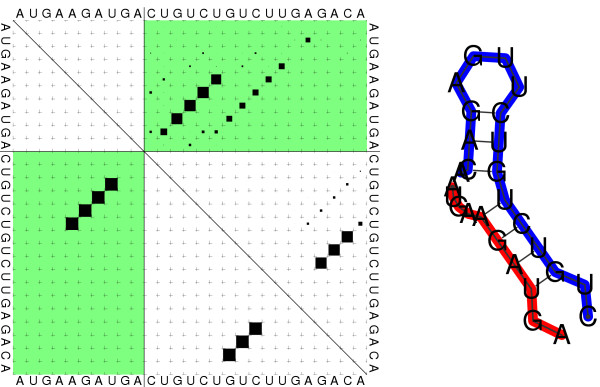
Results: We present a program, RNAcofold, that computes the hybridization energy and base pairing pattern of a pair of interacting RNA molecules. In contrast to earlier approaches, complex internal structures in both RNAs are fully taken into account. RNAcofold supports the calculation of the minimum energy structure and of a complete set of suboptimal structures in an energy band above the ground state. Furthermore, it provides an extension of McCaskill's partition function algorithm to compute base pairing probabilities, realistic interaction energies, and equilibrium concentrations of duplex structures.


Figures

Figure 1
Figure 1
Loops with cuts have to be scored differently. Top row: hairpins and interior loops containing the cut between n1 (black ball) and n1 + 1 (white ball). Below: multi loops containing the cut. Neither M1 nor M components must start at n1 + 1 or stop at n1. Note that the construction of ZM out of ZM and ZM1 ensures that the cut is not inside the loop part of ZM either.
Figure 2
Figure 2
Dot plot (left) and mfe structure representation (right) of the cofolding structure of the two RNA molecules AUGAAGAUGA (red) and CUGUCUGUCUUGAGACA (blue). Dot Plot: Upper right: Partition function. The area of the squares is proportional to the corresponding pair probabilities. Lower left: Minimum free energy structure. The two lines forming a cross indicate the cut point, intermolecular base pairs are depicted in the green upper right (partition function) and lower left (mfe) rectangle.
Figure 3
Figure 3
Example for the concentration dependency for two mRNA-siRNA binding experiments. In [54], Schubert et al. designed several mRNAs with identical target sites for an siRNA si, which are located in different secondary structures. In variant A, the VR1 straight mRNA, the binding site is unpaired, while in the mutant mRNA VR1 HP5-11, A', only 11 bases remain unpaired. We assume an mRNA concentration of a = 10 nmol/1 for both experiments. Despite the similar binding pattern, the binding energies (ΔF = FAB - FA - FB) differ dramatically. In [54], the authors observed 10% expression for VR1 straight, and 30% expression for the HP5-11 mutant. Our calculation shows that even if siRNA is added in excess, a large fraction of the VR1 HP5-11 mRNA remains unbound.
Figure 4
Figure 4
Difference dot Plot of native and mutated secondary structure of a 3 GU mutation of the CXCR4 siRNA gene. The red part on the right hand side shows the base pairing probability of the 5' part of the micro RNA, which is 80% higher in the native structure. This is an alternative explanation for the missing function of the mutant. Because of the mutations, the stack a little to the left gets more stable, and the probability of binding of the 5' end of the siRNA is reduced significantly.The color of the dots encodes the difference of the pair probabilities in the two molecules such positive (red) squares denote pairs more more probable in the second molecule (see color bar). The area of the dots is proportional to the larger of the two pair probabilities.

References

    1. Ambros V. The functions of animal microRNAs. Nature. 2004;431:350–355. doi: 10.1038/nature02871. - DOI - PubMed
    1. Kidner CA, Martienssen RA. The developmental role of microRNA in plants. Curr Opin Plant Biol. 2005;8:38–44. doi: 10.1016/j.pbi.2004.11.008. - DOI - PubMed
    1. Rivas E, Klein RJ, Jones TA, Eddy SR. Computational identification of noncoding RNAs in E. coli by comparative genomics. Curr Biol. 2001;11:1369–1373. doi: 10.1016/S0960-9822(01)00401-8. - DOI - PubMed
    1. McCutcheon JP, Eddy SR. Computational identification of non-coding RNAs in Saccharomyces cerevisiae by comparative genomics. Nucl Acids Res. 2003;31:4119–4128. doi: 10.1093/nar/gkg438. - DOI - PMC - PubMed
    1. Klein RJ, Misulovin Z, Eddy SR. Noncoding RNA genes identified in AT-rich hyperthermophiles. Proc Natl Acad Sci USA. 2002;99:7542–7547. doi: 10.1073/pnas.112063799. - DOI - PMC - PubMed

LinkOut - more resources