

Development of an experimental model of infected skin ulcer
- PMID: 16722897
- PMCID: PMC7951774
- DOI: 10.1111/j.1742-481x.2004.00006.x
Development of an experimental model of infected skin ulcer
Abstract
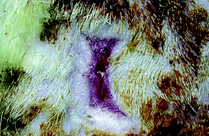
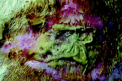
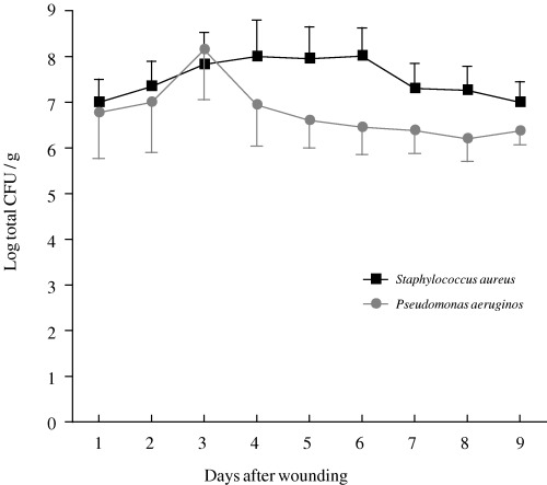
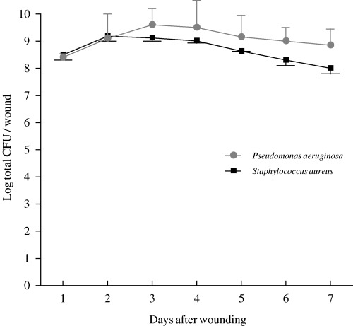
A model of infected skin ulceration could prove useful in assessing the clinical effectiveness of antimicrobial ointments and dressings. However, no such models have been previously established. Three types of wound were induced in rats: full-thickness wounds covered with gauze, burn wounds and wounds resulting from mechanical trauma. Wounds were inoculated with S. aureus or P. aeruginosa. Persistent infected wounds were observed only in full-thickness wounds covered with gauze. In a second experiment, colonies of P. aeruginosa or S. aureus were counted within 15 x 15 mm full-thickness wounds covered with gauze. Wounds were inoculated with 1.0 x 10(6) colony-forming units (CFU) of P. aeruginosa or S. aureus and then sealed to ensure an enclosed environment. Tissue bacterial counts exceeded 10(6) CFU/g from the next day until day 9 after infection. Bacterial counts exceeded 10(8) CFU/ml in wound exudate collected between days 1 and 7. We have developed a model of wound infection in which persistence of infection can be achieved for 9 days following ulceration due to the application of gauze to the base of a full-thickness wound.
Figures
References
-
- Schultz GS, Sibbald RG, Falanga V, Ayello EA, Dowsett C, Harding K, Romanelli M, Stacey MC, Teot L, Vanscheidt W. Wound bed preparation: a systematic approach to wound management. Wound Repair Regen 2003;11 Suppl: S1–28. - PubMed
-
- Bowler PG. Wound pathophysiology, infection and therapeutic options. Ann Med 2002;34: 419–27. - PubMed
-
- Robson MC. Wound infection. A failure of wound healing caused by an imbalance of bacteria. Surg Clin North Am 1997;77: 637–50. - PubMed
MeSH terms
LinkOut - more resources
Full Text Sources
