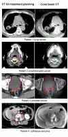
Correction of patient positioning errors based on in-line cone beam CTs: clinical implementation and first experiences
- PMID: 16723023
- PMCID: PMC1557518
- DOI: 10.1186/1748-717X-1-16
Correction of patient positioning errors based on in-line cone beam CTs: clinical implementation and first experiences
Abstract
Background: The purpose of the study was the clinical implementation of a kV cone beam CT (CBCT) for setup correction in radiotherapy.
Patients and methods: For evaluation of the setup correction workflow, six tumor patients (lung cancer, sacral chordoma, head-and-neck and paraspinal tumor, and two prostate cancer patients) were selected. All patients were treated with fractionated stereotactic radiotherapy, five of them with intensity modulated radiotherapy (IMRT). For patient fixation, a scotch cast body frame or a vacuum pillow, each in combination with a scotch cast head mask, were used. The imaging equipment, consisting of an x-ray tube and a flat panel imager (FPI), was attached to a Siemens linear accelerator according to the in-line approach, i.e. with the imaging beam mounted opposite to the treatment beam sharing the same isocenter. For dose delivery, the treatment beam has to traverse the FPI which is mounted in the accessory tray below the multi-leaf collimator. For each patient, a predefined number of imaging projections over a range of at least 200 degrees were acquired. The fast reconstruction of the 3D-CBCT dataset was done with an implementation of the Feldkamp-David-Kress (FDK) algorithm. For the registration of the treatment planning CT with the acquired CBCT, an automatic mutual information matcher and manual matching was used.
Results and discussion: Bony landmarks were easily detected and the table shifts for correction of setup deviations could be automatically calculated in all cases. The image quality was sufficient for a visual comparison of the desired target point with the isocenter visible on the CBCT. Soft tissue contrast was problematic for the prostate of an obese patient, but good in the lung tumor case. The detected maximum setup deviation was 3 mm for patients fixated with the body frame, and 6 mm for patients positioned in the vacuum pillow. Using an action level of 2 mm translational error, a target point correction was carried out in 4 cases. The additional workload of the described workflow compared to a normal treatment fraction led to an extra time of about 10-12 minutes, which can be further reduced by streamlining the different steps.
Conclusion: The cone beam CT attached to a LINAC allows the acquisition of a CT scan of the patient in treatment position directly before treatment. Its image quality is sufficient for determining target point correction vectors. With the presented workflow, a target point correction within a clinically reasonable time frame is possible. This increases the treatment precision, and potentially the complex patient fixation techniques will become dispensable.
Figures


References
MeSH terms
LinkOut - more resources
Full Text Sources
Other Literature Sources
Research Materials
Miscellaneous

