. 2006 May 24:3:34.
doi: 10.1186/1743-422X-3-34.

Contribution of cysteine residues in the extracellular domain of the F protein of human respiratory syncytial virus to its function

Affiliations

Contribution of cysteine residues in the extracellular domain of the F protein of human respiratory syncytial virus to its function

Nicole D Day et al. Virol J. .

Abstract

The mature F protein of all known isolates of human respiratory syncytial virus (HRSV) contains fifteen absolutely conserved cysteine (C) residues that are highly conserved among the F proteins of other pneumoviruses as well as the paramyxoviruses. To explore the contribution of the cysteines in the extracellular domain to the fusion activity of HRSV F protein, each cysteine was changed to serine. Mutation of cysteines 37, 313, 322, 333, 343, 358, 367, 393, 416, and 439 abolished or greatly reduced cell surface expression suggesting these residues are critical for proper protein folding and transport to the cell surface. As expected, the fusion activity of these mutations was greatly reduced or abolished. Mutation of cysteine residues 212, 382, and 422 had little to no effect upon cell surface expression or fusion activity at 32 degrees C, 37 degrees C, or 39.5 degrees C. Mutation of C37 and C69 in the F2 subunit either abolished or reduced cell surface expression by 75% respectively. None of the mutations displayed a temperature sensitive phenotype.


Figures

Figure 1
Figure 1
Diagram of the HRSV F protein. A linear representation of the HRSV F precursor protein (A2 strain) is shown. Amino acid positions of individual domains are indicated with residues numbered in the context of the full-length coding region. Disulfide linked F1 & F2 subunits are delineated with arrows. The furin mediated cleavage sites are indicated by filed arrowheads. The intervening cleavage fragment is indicated as a gray box. Positions of the individual cysteine residues are depicted as asterisks. Asparagine residues (N116 and N126) which are sites of N-linked glycosylation are represented with circles. The site of palmitoylation at cysteine residue 550 is depicted as a jagged line. SP = signal peptide; f = fusion peptide; HR1 = heptad repeat 1; HR2 = heptad repeat 2; TM = transmembrane region. Figure adapted from [5].
Figure 2
Figure 2
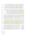
Alignment of paramyxoviral F proteins. Sequence alignment was performed as described in methods. Accession numbers for the sequences of the viral F proteins used for the alignment are as described in methods. Conserved cysteine residues are highlighted in yellow.
Figure 3
Figure 3
Computer model of the HRSV F protein. The molecular structure of HRSV F protein ECD was modeled using the human parainfluenza virus 3 virus F protein ECD structure as template as described in methods. Ribbon diagrams of the F1-F2 monomer (left) and F protein homotrimer (right) are shown. Heptad repeat 1 (HR1) and heptad repeat 2 (HR2) are indicated with arrows. Cysteine residues are depicted as yellow balls with specific residue disulfide pairs indicated on the monomer.
Figure 4
Figure 4
Immunoprecipitation of HRSV F cysteine mutations. 293T cells were mock transfected (-), transfected with a plasmid expressing beta-galactosidase (b-gal), or plasmids encoding the wild-type (WT) HRSV F protein or various cysteine mutants (listed above lanes), followed by metabolic labeling with [35S]-methionine/cysteine mixture, and immunoprecipitation as described in [44]. The positions of molecular weight size markers are indicated. The positions of the F1 and F2 subunits are indicated with arrows.
Figure 5
Figure 5
Expression of cysteine mutations measured by ELISA. 293T cells were transfected with plasmids encoding the wild-type HRSV F (WT), empty vector cassette (EV) or the various cysteine mutants (listed below lanes), followed by fixation and analysis using an ELISA as described in methods. Results are presented relative to values obtained with wild-type HRSV F which was set at 100%, and represent the average of three separate determinations. Results obtained using permeabilizing conditions are depicted with open bars. Results obtained using non-permeabilizing conditions are depicted with a solid bars.
Figure 6
Figure 6
Fusion activity of cysteine mutations. 293T cells were transfected with plasmids encoding either the wild-type HRSV F protein or the panel of cysteine mutants and fusion activity was measured at 32°C, 37°C, or 39.5°C as described in [44]. Fusion activity is represented as relative light units (RLUs), and values represent the average of three separate determinations.

References

    1. Han LL, Alexander JP, Anderson LJ. Respiratory syncytial virus pneumonia among the elderly: an assessment of disease burden. J Infect Dis. 1999;179:25–30. doi: 10.1086/314567. - DOI - PubMed
    1. Ison MG, Hayden FG. Viral infections in immunocompromised patients: what's new with respiratory viruses? Curr Opin Infect Dis. 2002;15:355–367. - PubMed
    1. Thompson WW, Shay DK, Weintraub E, Brammer L, Cox N, Anderson LJ, Fukuda K. Mortality associated with influenza and respiratory syncytial virus in the United States. JAMA. 2003;289:179–186. doi: 10.1001/jama.289.2.179. - DOI - PubMed
    1. Whimbey E, Ghosh S. Respiratory syncytial virus infections in immunocompromised adults. Curr Clin Top Infect Dis. 2000;20:232–255. - PubMed
    1. Collins PL, Chanock RM, Murphy BR. Respiratory syncytial virus. In: Knipe DM and Howley PM, editor. Fields Virology. Fourth. Vol. 1. Philadelphia, Lippincott, Williams, and Wilkins; 2001. pp. 1443–1485.