Clinical Trial
. 2006 May 24;26(21):5777-85.
doi: 10.1523/JNEUROSCI.5223-05.2006.

Pronociceptive and antinociceptive effects of estradiol through endogenous opioid neurotransmission in women

Affiliations
Clinical Trial

Pronociceptive and antinociceptive effects of estradiol through endogenous opioid neurotransmission in women

Yolanda R Smith et al. J Neurosci. .

Abstract

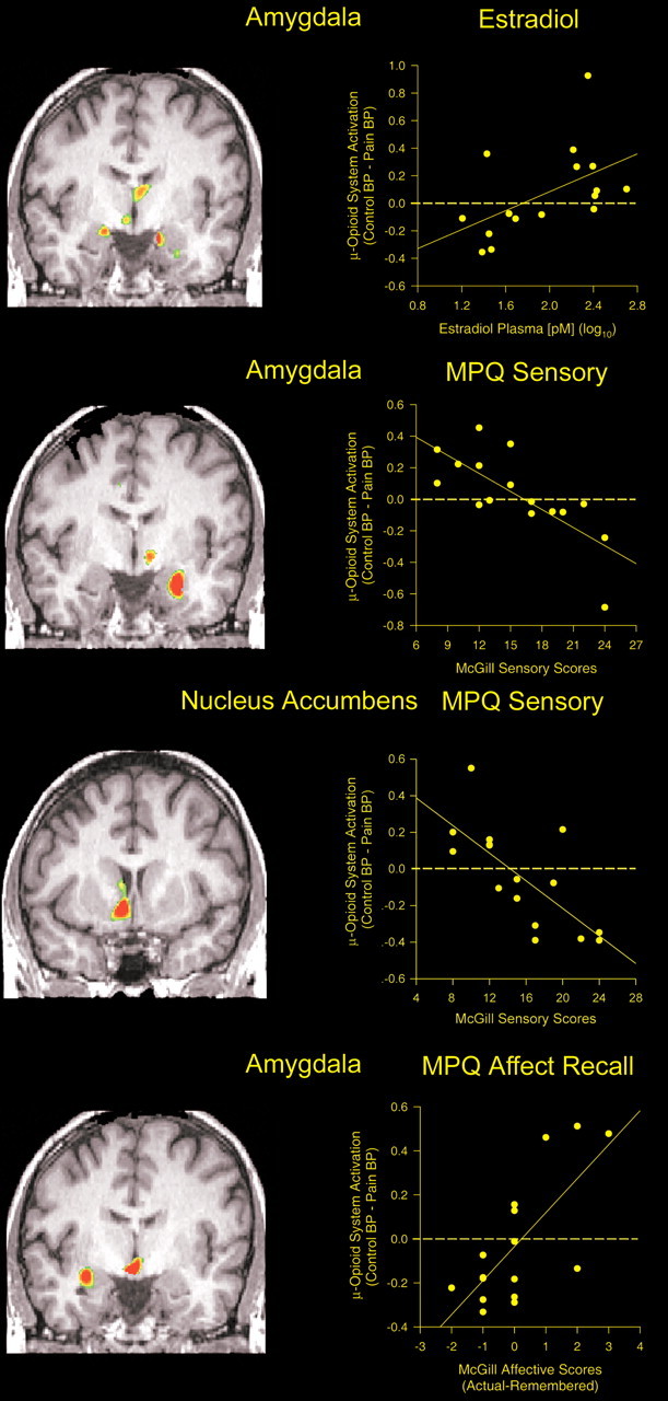
Prominent interindividual and sex-dependent differences have been described in responses to sustained pain and other stressful stimuli. Variations in mu-opioid receptor-mediated endogenous opioid neurotransmission may underlie some of these processes. We examined both baseline mu-opioid receptor levels and the activation of this neurotransmitter system during sustained pain using positron emission tomography in a sample of young healthy men and women. Women were studied twice, during low and high estrogen states. The high-estrogen state was associated with regional increases in baseline mu-opioid receptor availability in vivo and a greater activation of endogenous opioid neurotransmission during the pain stressor. The latter did not differ from that obtained in males. During the low estrogen condition, however, significant reductions in endogenous opioid tone were observed at the level of thalamus, nucleus accumbens, and amygdala, which were associated with hyperalgesic responses. Estrogen-associated variations in the activity of mu-opioid neurotransmission correlated with individual ratings of the sensory and affective perceptions of the pain and the subsequent recall of that experience. These data demonstrate a significant role of estrogen in modulating endogenous opioid neurotransmission and associated psychophysical responses to a pain stressor in humans.


Figures

Figure 1.
Figure 1.
Effect of estradiol on μ-opioid receptor binding and on responses of μ-opioid receptor-mediated neurotransmission to a stress challenge. Top, left, Three-dimensional representation of μ-opioid receptor binding potential values in a representative healthy volunteer, superimposed over an MRI image standardized to ICBM stereotactic coordinates. Binding potential values are represented by the pseudocolor scale in the lower part of the figure. Top, center, Three-dimensional display of significant effects of estradiol treatment on baseline μ-opioid receptor binding potential for the entire sample, superimposed over a magnetic resonance image in ICBM stereotactic coordinates. Standardized z scores are represented by the pseudocolor scale on the left. Significant effects of estradiol treatment were observed in the medial thalamus, ventral basal ganglia, amygdala, and hypothalamus. The graph shows the mean ± SEM of BP values for thalamus, ventral basal ganglia, and amygdala during low and high estradiol states (yellow). BP values for a matched sample of eight men are shown in blue for demonstrative purposes. Bottom, Brain areas in which significant effects of estradiol were observed on stress-induced activation of the μ-opioid system for the entire sample. Standardized z scores are represented by the pseudocolor scale on the left. Significant effects of estradiol condition were observed in the medial thalamus, ventral basal ganglia, and amygdala. The graph shows the mean ± SEM of the change in BP for these regions during the stress challenge (negative values reflecting reductions in μ-opioid neurotransmission, positive values reflecting activation of neurotransmission) during low- and high-estradiol states (yellow). The change in BP values for a matched sample of eight men are shown in blue for demonstrative purposes.
Figure 2.
Figure 2.
Correlations between the stress-induced changes in μ-opioid neurotransmission, estradiol plasma levels, and other psychophysical measures. Top, Significant correlations between estradiol plasma levels in picomolar (log10) and stress-induced μ-opioid system responses were obtained for the medial thalamus, amygdala (shown), and the ventral basal ganglia (data not shown). A coronal view is shown at the y (anterior–posterior) coordinate, −5. The graph inset shows the individual values for this correlation in the amygdala. Second panel, Significant voxel-by-voxel correlations between stress-induced μ-opioid system responses and MPQ pain sensory scores as rated by the volunteers at the level of the amygdala (coronal view, y = −6). The graph shows the individual values for this correlation. Third panel, Significant voxel-by-voxel correlations between stress-induced μ-opioid system responses and MPQ pain sensory scores as rated by the volunteers at the level of the nucleus accumbens (coronal view, y = 5). The graph shows the individual values for this correlation. Bottom, Correlations between stress-induced μ-opioid system responses and the subsequent recall of the pain experience 24 h after the pain challenge. Significant effects of μ-opioid system activation on the recall of MPQ pain sensory scores were observed in the medial thalamus (data not shown). Effects on the recall of MPQ pain affect scores were detected in the amygdala, medial thalamus, and hypothalamus. The graph shows the individual values for the latter correlation in the amygdala (coronal view, y = −6).

References

    1. Akil H, Watson S, Young E, Lewis M, Khachaturian H, Walker J (1984). Endogenous opioids: biology and function. Annu Rev Neurosci 7:223–255. - PubMed
    1. Anderson AK, Christoff K, Stappen I, Panitz D, Ghahremani DG, Glover G, Gabrieli JD, Sobel N (2003). Dissociated neural representations of intensity and valence in human olfaction. Nat Neurosci 6:196–202. - PubMed
    1. Bond C, LaForge K, Tian M, Melia D, Zhang S, Borg L, Gong J, Schuler J, Strong J, Leal S, Tischfield J, Kreek M, Yu L (1998). Single-nucleotide polymorphism in the human mu opioid receptor gene alters β-endorphin binding and activity: possible implications for opiate addiction. Proc Natl Acad Sci USA 91:9081–9085. - PMC - PubMed
    1. Bonen A (1994). Exercise-induced menstrual cycle changes. A functional, temporary adaptation to metabolic stress. Sports Med 17:373–392. - PubMed
    1. Borras MC, Becerra L, Ploghaus A, Gostic JM, DaSilva A, Gonzalez RG, Borsook D (2004). fMRI measurement of CNS responses to naloxone infusion and subsequent mild noxious thermal stimuli in healthy volunteers. J Neurophysiol 91:2723–2733. - PubMed

Publication types