. 2006 May 24;26(21):5849-59.
doi: 10.1523/JNEUROSCI.4921-05.2006.

WAVE1 is required for oligodendrocyte morphogenesis and normal CNS myelination

Affiliations

WAVE1 is required for oligodendrocyte morphogenesis and normal CNS myelination

Hyun-Ju Kim et al. J Neurosci. .

Abstract

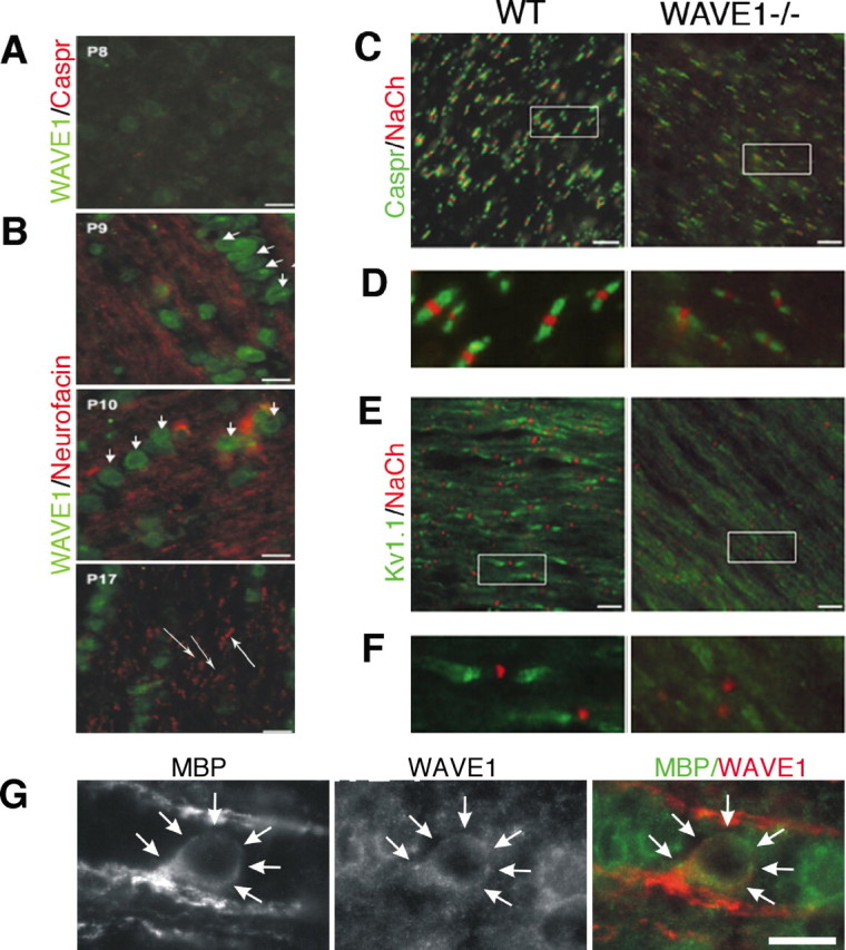
Myelin formation involves the outgrowth of an oligodendrocyte cell process that can be regarded as a giant lamellipodium because it is an actively growing structure with extruded cytoplasm. The actin cytoskeleton is critical to morphogenesis, but little is known about regulation of actin dynamics in oligodendrocytes. Wiskott-Aldrich syndrome protein family verprolin homologous (WAVE) proteins mediate lamellipodia formation; thus, we asked whether these proteins function in oligodendrocyte process formation and myelination. Here, we show that WAVE1 is expressed by oligodendrocytes and localizes to the lamella leading edge where actin polymerization is actively regulated. CNS WAVE1 expression increases at the onset of myelination. Expression of dominant-negative WAVE1 impaired process outgrowth and lamellipodia formation in cultured oligodendrocytes. Similarly, oligodendrocytes isolated from mice lacking WAVE1 had fewer processes compared with controls, whereas neurons and astrocytes exhibited normal morphology. In white matter of WAVE1-/- mice, we found regional hypomyelination in the corpus callosum and to a lesser extent in the optic nerve. In optic nerve from WAVE1-/- mice, there were fewer nodes of Ranvier but nodal morphology was normal, implicating a defect in myelin formation. Our in vitro findings support a developmentally dynamic and cell-autonomous role for WAVE1 in regulating process formation in oligodendrocytes. Additionally, WAVE1 function during CNS myelination appears to be linked to regional cues. Although its loss can be compensated for in many CNS regions, WAVE1 is clearly required for normal amounts of myelin to form in corpus callosum and optic nerve. Together, these data demonstrate a role for WAVE1 in oligodendrocyte morphogenesis and myelination.


Figures

Figure 1.
Figure 1.
WASP family protein expression pattern in mouse tissues. A, WASP (500 bp), N-WASP (390 bp), and WAVE1 (500 bp) transcripts were detected by RT-PCR in mouse tissues. B, Relative expression levels of WAVE1 protein (85 kDa) was detected by Western blot among the tissues indicated. C, Developmental expression profile of WAVE1 was determined by Western blot in protein extracts from E13 through adult mouse brain, showing that WAVE1 is expressed at E13 with triple bands, increased in developing brain, and peaked at P7, and reduced to a single band in adult brain. In contrast, WAVE2 (85 kDa) is expressed in all examined ages and WAVE3 (60 kDa) is steadily increased in developing brain until adult. D, Cell type distributions of WAVE1 by Western blot using extracts of purified cell types revealed strong expression in neurons and oligodendrocytes, and low levels of expression in astrocytes and microglia. In contrast, WAVE2 is expressed in all cell types examined and WAVE3 in neurons only. β-Actin primer (350 bp) was used for transcription level of actin as a positive control in A, and anti-actin monoclonal antibody (42 kDa) was used for a loading control (B–D). M, Molecular weight marker. E, Wave1 expression was assessed by in situ and by LacZ staining. The left side of E shows in situ signaling from coronal brain slices of normal 129/svLex mice at ∼29 d of age. On the right side, the lacZ staining of Wave KO mice at ∼21 d of age illustrates the general persistence of cortical and subcortical LacZ+ cells, whose wave1 locus is transcriptionally active. The bottom two panels are parasagittal sections of adult WT mice with WAVE1 antisense (left) and sense (right) controls.
Figure 2.
Figure 2.
Cloning and generation of vectors for DN-WAVE1. A, Diagrammatic representations of the domain structure of rat WAVE1 gene with the structures and names of the chimeric constructs are shown. WAVE1 contains an N-terminal SHD, a proline-rich internal segment (PRO), and a C-terminal VCA domain. B, The WAVE1 constructs were cloned to pEGFPC3 (Clontech), and proteins were expressed in HEK293 cells. Their size was determined by Western blot using a monoclonal antibody against GFP or WAVE1. Arrows in B represent the expected size of the GFP-fusion proteins. C, WAVE1 adenoviral constructs were generated to transfect primary cells and transduced into purified oligodendrocytes isolated from P2 rat and cortical neurons isolated from E17 mice. Infected cells were then grown for 48 h, fixed, and analyzed. Overexpression of dWA and dA blocked process formation in oligodendrocytes, but no effect was seen in neurons. D, Higher magnification of insets in C is seen in D. E, Oligodendrocyte processes were counted in two nonoverlapping fields, and experiments were performed in four independent experiments. F, TUNEL assay shows that oligodendrocyte death in response to viral transduction was not significant compared with GFP alone. Error bars indicate SEM. ∗∗p < 0.001 and ∗p < 0.005 by ANOVA (n = 4). Scale bars, 10 μm. G, Oligodendrocytes transfected with GFP-dWA and GFP-vector alone were also stained with rhodamine phalloidin (red) and used for measurements in lamellipodial size. Note that elaborate lamellipodia at the end of oligodendrocytes process in GFP-control (arrows) compared with GFP-dWA-transfected cells (arrowheads).
Figure 3.
Figure 3.
WAVE1 defect causes delayed oligodendrocyte differentiation. Mixed CNS cells from P0 WT and WAVE−/− mice brain were dissociated in DMEM and cultured for 15 d with changing medium every 3 d. A, Neurofilament (NF), glial fibrillary acidic protein (GFAP), and O4 were stained for different populations of CNS cells. Oligodendrocytes were double labeled with O4/Olig2 (B) or O1/Olig2 (C). Significant defects in process formation were seen in WAVE−/− O4+ cells compared with WT controls. D, Total cell number by DAPI+ and total oligodendrocyte number by Olig2+ were counted in square millimeters. Cell numbers counted WT versus WAVE−/− was not statistically significant. E, However, the number of O4+/Olig2+ and O1+/Olig2+ double-stained cells was significantly reduced. In addition, mature O1+ oligodendrocytes with membrane sheets were dramatically reduced (72%) compared with wild-type control (F). The data shown here are representative of four independent experiments. Error bars indicate SEM. ∗p < 0.05 and ∗∗p < 0.005 by ANOVA (n = 4). Scale bars: A, 20 μm; B, 5 μm; C, 10 μm.
Figure 4.
Figure 4.
Subcellular localization of WAVE1 in vitro. Fluorescence images of WAVE1 localization in oligodendrocytes purified from the forebrains of 2-d-old rats. A, WAVE1 (red) staining shows a dot-like continuous localization (arrows) along the leading edge of lamellipodia in early stage of OPCs. Higher magnification inset is seen in the right panel of A. F-actin (green) was labeled with Alexa Fluor 488 phalloidin. B, WAVE1 (red) is concentrated in the protruding tips of A2B5+ (green) oligodendrocytes, distributed along the processes of O4+, O1+, and MBP+ (green) mature oligodendrocytes (long arrows), and cytoplasm of cell bodies. Note that WAVE1 staining is localized at the leading edge of membrane sheet in O1+ cells (short arrows). Scale bars: A, 30 μm; B, 30 μm.
Figure 5.
Figure 5.
Clustering of Na+ and K+ channels at and near nodes is decreased in developing WAVE1−/− mouse optic nerve. A, Immunohistochemistry of P8 rat optic nerve revealed weak expression of WAVE1 and no expression of Caspr (red), a specific marker of paranodes. B, Increased expression of WAVE1 (green) is seen in P9 through P17 rat optic nerve. Expression of WAVE1 is mainly restricted to oligodendrocyte cytoplasm (short arrows). Neurofascin (red), a specific marker for nodes and paranodes, is evident in developing optic nerve (long arrows). C, P21 mouse optic nerve was double labeled with NaCh to mark the node and Caspr to mark the paranode and staining with NaCh and Kv1.1 to mark the juxtaparanode (E). Higher magnification of insets in C and E is seen in D and F. Nodal structure is normal in WAVE1−/−, but fewer nodes are seen. G, High-power image of an oligodendrocyte within the optic nerve double labeled for MBP and WAVE1. Scale bars, 10 μm.
Figure 6.
Figure 6.
The corpus callosum is thinner and the total number of oligodendrocytes is reduced in WAVE1−/−. A, CC1, a marker for mature oligodendrocytes, was immunostained with coronal sections of P21 mouse brain. B, Twenty-micrometer-thick coronal sections of P20 mouse brain were stained with a specific marker of pan-oligodendrocytes, Olig2, and detected by the ABC Elite kit with DAB substrate. Higher magnification is seen in the bottom panel. Fewer CC1+ cells and Olig2+ cells are obvious in corpus callosum (CC) of WAVE1−/− compared with WT controls. C, The number of Olig2+ cells was quantified within the designated callosal region in black line with two consecutive sections. Although the number of Olg2+ in CC is reduced (B), the density per square millimeter of Olig2+ cells is slightly increased in most areas except the corpus callosum, in which the cell density is virtually identical to WT. Interestingly, Olig2+ cells per square millimeter of cortex in WAVE1−/− were higher than WT by ∼53%. Error bars indicate SEM. ∗p < 0.05 (n = 6) and ∗∗p < 0.001 [n = 6 for CC and n = 4 for cortex (CTX), striatum (ST), septum (SEP)] by ANOVA. Scale bar, 10 μm. Arrows in A and B indicate inter-hemispheric fissure of the coronal section.
Figure 7.
Figure 7.
WAVE1−/− mice reveal defective myelination in the CNS, with severe hypomyelination seen in the corpus callosum. A, Electron micrographs of the midsagittal corpus callosum (CC) sections were analyzed from WAVE1−/− mice at P20 as well as WT littermates. Axons in WAVE1−/− mice are hypomyelinated, and fewer total myelinated axons are clearly seen. The majority of myelin sheaths appears properly compacted, but shown is a rare sheath that is poorly compacted (arrows). The primary disturbance appears to be that many axons are unmyelinated (arrowheads) in WAVE1−/− compared with WT. B, C, Cross sections of the middle portion of the eyeball and optic chiasm of optic nerve (ON) and the cervical region of the spinal cord (SP) were photographed by electron microscope. No prominent differences were observed in those areas except that the number of axons was reduced slightly in ON. D, The total number of myelinated axons in midsagittal corpus callosum was counted with three different P20 WAVE1−/− animal and two different P20 WT littermate controls. We counted myelinated axons of ON and SP in square millimeters of eight fields of view for WAVE−/− and six fields for WT from three different P20 animals for each genotype. Error bars indicate SEM. ∗p < 0.05 and ∗∗p < 0.005 by ANOVA, respectively. Scale bars: A, 200 nm; B, 500 nm; C, 1 μm.

References

    1. Asou H, Hamada K, Sakota T (1995). Visualization of a single myelination process of an oligodendrocyte in culture by video microscopy. Cell Struct Funct 20:59–70. - PubMed
    1. Baron W, Colognato H, ffrench-Constant C (2005). Integrin-growth factor interactions as regulators of oligodendroglial development and function. Glia 49:467–479. - PubMed
    1. Barres BA, Raff MC (1994). Control of oligodendrocyte number in the developing rat optic nerve. Neuron 12:935–942. - PubMed
    1. Bjartmar C, Kinkel RP, Kidd G, Rudick RA, Trapp BD (2001). Axonal loss in normal-appearing white matter in a patient with acute MS. Neurology 57:1248–1252. - PubMed
    1. Bompard G, Caron E (2004). Regulation of WASP/WAVE proteins: making a long story short. J Cell Biol 166:957–962. - PMC - PubMed

Publication types

Substances