Comparative Study
. 2006 Jun;50(6):2156-66.
doi: 10.1128/AAC.01499-05.

Functional, biophysical, and structural bases for antibacterial activity of tigecycline

Affiliations
Comparative Study

Functional, biophysical, and structural bases for antibacterial activity of tigecycline

Matthew W Olson et al. Antimicrob Agents Chemother. 2006 Jun.

Abstract

Tigecycline is a novel glycylcycline antibiotic that possesses broad-spectrum activity against many clinically relevant species of bacterial pathogens. The mechanism of action of tigecycline was delineated using functional, biophysical, and molecular modeling experiments in this study. Functional assays showed that tigecycline specifically inhibits bacterial protein synthesis with potency 3- and 20-fold greater than that of minocycline and tetracycline, respectively. Biophysical analyses demonstrated that isolated ribosomes bind tigecycline, minocycline, and tetracycline with dissociation constant values of 10(-8), 10(-7), and >10(-6) M, respectively. A molecular model of tigecycline bound to the ribosome was generated with the aid of a 3.40-angstrom resolution X-ray diffraction structure of the 30S ribosomal subunit from Thermus thermophilus. This model places tigecycline in the A site of the 30S subunit and involves substantial interactions with residues of H34 of the ribosomal subunit. These interactions were not observed in a model of tetracycline binding. Modeling data were consistent with the biochemical and biophysical data generated in this and other recent studies and suggested that tigecycline binds to bacterial ribosomes in a novel way that allows it to overcome tetracycline resistance due to ribosomal protection.


Figures

FIG. 1.
FIG. 1.
Structure of tigecycline.
FIG. 2.
FIG. 2.
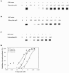
Effect of tigecycline, tetracycline, and minocycline on protein synthesis. IVT reactions were performed in the presence of tigecycline (A), minocycline (B), or tetracycline (C). Compounds were added as indicated prior to the addition of GFP plasmid vector. Samples were separated by sodium dodecyl sulfate-polyacrylamide gel electrophoresis, and labeled protein was detected using a phosphorimager. Bands represent the 35S-labeled GFP (∼28 kDa). (D) IC50 value analysis from the data in panels A to C.
FIG. 3.
FIG. 3.
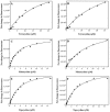
Binding isotherms for the interactions of tigecycline, minocycline, and tetracycline with 30S and 70S ribosomes where compound is titrated into the system. The isolated 30S (A, C, and E) and 70S (B, D, and F) ribosomes were incubated with tetracycline (A and B), minocycline (C and D), and tigecycline (E and F). Samples contained 1.5 μM of ribosomes for tetracycline and 0.5 μM for tigecycline and minocycline. The net change in fluorescence for tigecycline, minocycline, and tetracycline was measured at 515 to 520 nm, 505 to 510 nm, and 515 to 520 nm, respectively, in the presence of increasing concentrations of compound: 0 to 25 μM tetracycline, 0 to 15 μM tigecycline, and 0 to 15 μM minocycline. The solid line in each panel corresponds to the curve fit to the quadratic equation.
FIG. 4.
FIG. 4.
Binding isotherms for the interactions of tigecycline, minocycline, and tetracycline with 30S and 70S ribosomes where ribosome is titrated into the system. Tigecycline (A and B), minocycline (C and D), and tetracycline (E and F) were present at 1 μM concentrations whereby the isolated 30S (A, C, and E) and 70S (B, D, and F) ribosomes were titrated into the system. The net change in fluorescence for tigecycline, minocycline, and tetracycline was measured at 510 to 520 nm, 505 to 515 nm, and 510 to 520 nm, respectively, in the presence of increasing concentrations of ribosome. The solid line in each panel corresponds to the curve fit to the quadratic equation.
FIG. 5.
FIG. 5.
Competition of [3H]tetracycline and [14C]tigecycline by unlabeled tigecycline, minocycline, and tetracycline. The 30S (40 pmol, 0.6 μM [A and C]) and 70S (70 pmol, 1.4 μM [B and D]) ribosomes were incubated for 15 min at 37°C with [3H]tetracycline (6 μM [A and B]) or [14C]tigecycline (4 μM [C and D]) in the presence of increasing concentrations of unlabeled tigecycline, minocycline, and tetracycline (0 to 200 μM). The quantity of [3H]tetracycline and [14C]tigecycline bound to the ribosomes was determined. For panels A to D, 100% binding represents the amount of radiolabeled compound bound in the absence of unlabeled compound. For panel A these values are 4, 4.5, and 4.7 pmol of [3H]tetracycline for tigecycline, minocycline, and tetracycline titrated into the assay, respectively. Likewise for panel B these values are 4.2, 6.3, and 6.9 pmol, respectively. For panel C, the quantities of [14C]tigecycline bound in the absence of unlabeled compound are 25, 34, and 30 pmol for tigecycline, minocycline, and tetracycline titrated into the system, respectively. Similarly for panel D, these values are 50, 64, and 58 pmol, respectively.
FIG. 6.
FIG. 6.
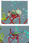
Computational docking. A. Overview of the tigecycline binding site and the predicted ligand-ribosome interactions. The RNA helices H34, H31, and H18 are colored in red, cyan, and yellow, respectively. B. Overview of the tetracycline-ribosome interaction and the predicted minocycline-ribosome interactions. The RNA helices H34, H31, and H18 are colored in red, cyan, and yellow, respectively. Tetracycline is represented with its carbon atoms colored in green and minocycline with its carbon atoms colored in pink. The green double-headed arrow represents the π-stacking interaction between the D ring and the cytosine of C1054.
FIG. 7.
FIG. 7.
Chemical structure diagram of the main interactions between tigecycline and the 16S RNA in the A binding site. The dashed lines represent the hydrogen bonds whereas the dotted lines represent the coordination bonds between tigecycline and a magnesium ion bound to the surface of the ribosome.

References

    1. Bauer, G., C. Berens, S. J. Projan, and W. Hillen. 2004. Comparison of tetracycline and tigecycline binding to ribosomes mapped by dimethylsulphate and drug-directed Fe2+ cleavage of 16S rRNA. J. Antimicrob. Chemother. 53:592-599. - PubMed
    1. Bergeron, J., M. Ammirati, D. Danley, L. James, M. Norcia, J. Retsema, C. A. Strick, W. G. Su, J. Sutcliffe, and L. Wondrack. 1996. Glycylcyclines bind to the high-affinity tetracycline ribosomal binding site and evade Tet(M)- and Tet(O)-mediated ribosomal protection. Antimicrob. Agents Chemother. 40:2226-2228. - PMC - PubMed
    1. Boucher, H. W., C. B. Wennersten, and G. M. Eliopoulos. 2000. In vitro activities of the glycylcycline GAR-936 against gram-positive bacteria. Antimicrob. Agents Chemother. 44:2225-2229. - PMC - PubMed
    1. Bouchillon, S. K., D. J. Hoban, B. M. Johnson, T. M. Stevens, M. J. Dowzicky, D. H. Wu, and P. A. Bradford. 2005. In vitro evaluation of tigecycline and comparative agents in 3049 clinical isolates: 2001 to 2002. Diagn. Microbiol. Infect. Dis. 51:291-295. - PubMed
    1. Bradford, P. A. 2004. Tigecycline: a first in class glycylcycline. Clin. Microbiol. Newsl. 26:163-168.

Publication types

MeSH terms