Diabetes enhances periodontal bone loss through enhanced resorption and diminished bone formation
- PMID: 16723646
- PMCID: PMC2253683
- DOI: 10.1177/154405910608500606
Diabetes enhances periodontal bone loss through enhanced resorption and diminished bone formation
Abstract
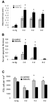
Using a ligature-induced model in type-2 Zucker diabetic fatty (ZDF) rat and normoglycemic littermates, we investigated whether diabetes primarily affects periodontitis by enhancing bone loss or by limiting osseous repair. Diabetes increased the intensity and duration of the inflammatory infiltrate (P < 0.05). The formation of osteoclasts and percent eroded bone after 7 days of ligature placement was similar, while four days after removal of ligatures, the type 2 diabetic group had significantly higher osteoclast numbers and activity (P < 0.05). The amount of new bone formation following resorption was 2.4- to 2.9-fold higher in normoglycemic vs. diabetic rats (P < 0.05). Diabetes also increased apoptosis and decreased the number of bone-lining cells, osteoblasts, and periodontal ligament fibroblasts (P < 0.05). Thus, diabetes caused a more persistent inflammatory response, greater loss of attachment and more alveolar bone resorption, and impaired new bone formation. The latter may be affected by increased apoptosis of bone-lining and PDL cells.
Figures


References
-
- Alikhani M, Alikhani Z, Graves DT. Apoptotic effects of LPS on fibroblasts are indirectly mediated through TNFR1. J Dent Res. 2004;83:671–676. - PubMed
-
- Alikhani Z, Alikhani M, Boyd CM, Nagao K, Trackman PC, Graves DT. Advanced glycation end products enhance expression of pro-apoptotic genes and stimulate fibroblast apoptosis through cytoplasmic and mitochondrial pathways. J Biol Chem. 2005;280:12087–12095. - PubMed
-
- Assuma R, Oates T, Cochran D, Amar S, Graves DT. IL-1 and TNF antagonists inhibit the inflammatory response and bone loss in experimental periodontitis. J Immunol. 1998;160:403–409. - PubMed
-
- Bacic M, Plancak D, Granic M. CPITN assessment of periodontal disease in diabetic patients. J Periodontol. 1988;59:816–822. - PubMed
-
- Bezerra MM, Brito GA, Ribeiro RA, Rocha FA. Low-dose doxycycline prevents inflammatory bone resorption in rats. Braz J Med Biol Res. 2002;35:613–616. - PubMed
Publication types
MeSH terms
Grants and funding
LinkOut - more resources
Full Text Sources
Other Literature Sources
Medical

