. 2006 Jun;168(6):1808-20.
doi: 10.2353/ajpath.2006.051091.

Loss of angiotensin-converting enzyme-2 leads to the late development of angiotensin II-dependent glomerulosclerosis

Affiliations

Loss of angiotensin-converting enzyme-2 leads to the late development of angiotensin II-dependent glomerulosclerosis

Gavin Y Oudit et al. Am J Pathol. 2006 Jun.

Abstract

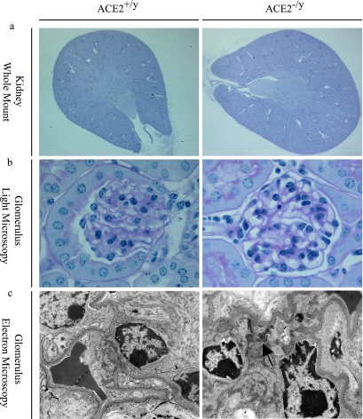
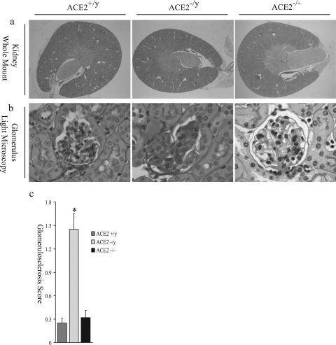
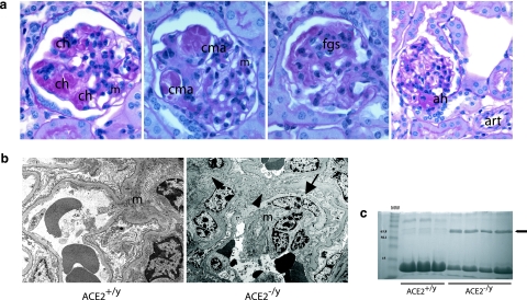
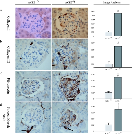
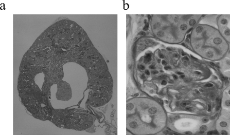
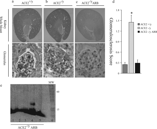
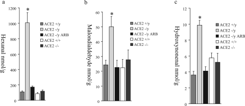
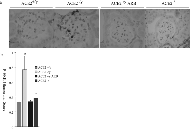
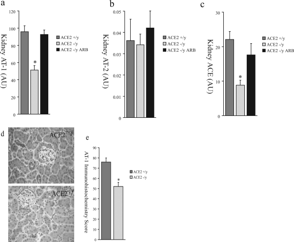
Angiotensin-converting enzyme-2 (ACE2), a membrane-bound carboxymonopeptidase highly expressed in the kidney, functions as a negative regulator of the renin-angiotensin system. Here we report early accumulation of fibrillar collagen in the glomerular mesangium of male ACE2 mutant (ACE2-/y) mice followed by development of glomerulosclerosis by 12 months of age whereas female ACE2 mutant (ACE2-/-) mice were relatively protected. Progressive kidney injury was associated with increased deposition of collagen I, collagen III and fibronectin in the glomeruli and increased urinary albumin excretion compared to age-matched control mice. These structural and functional changes in the glomeruli of male ACE2 mutant mice were prevented by treatment with the angiotensin II type-1 receptor antagonist irbesartan. Loss of ACE2 was associated with a marked increase in renal lipid peroxidation product formation and activation of mitogen-activated protein kinase and extracellular signal-regulated kinases 1 and 2 in glomeruli, events that are also prevented by angiotensin II type-1 receptor blockade. We conclude that deletion of the ACE2 gene leads to the development of angiotensin II-dependent glomerular injury in male mice. These findings have important implications for our understanding of ACE2, the renin-angiotensin system, and gender in renal injury, with ACE2 likely to be an important therapeutic target in kidney disease.


Figures

Figure 1
Figure 1
Normal architecture in young (3-month-old) ACE2 mutant mice with early ultrastructural changes. a: Whole mount of ACE2−/y mouse kidneys showing normal architecture (PAS, original magnification, ×16). b: ACE2−/y glomeruli are normal by light microscopy, comparable to ACE2+/y controls (PAS, original magnification, ×600). c: Electron microscopy shows early fibrillar collagen deposition (arrow) in the ACE2−/y mice but not in wild-type controls (original magnification, ×4800).
Figure 2
Figure 2
Glomerulosclerosis in old (1-year) ACE2 mutant mice. a: Whole mount of ACE2+/y, ACE2−/y, and ACE2−/− (female mutant mice) mouse kidneys show normal architecture (PAS, original magnification, ×16). b: ACE2−/y, but not ACE2+/yor ACE2−/−, glomeruli show glomerulosclerosis, segmental scarring, and hyalinosis (PAS, original magnification, ×600). c: Glomerulosclerosis scores of ACE2+/y, ACE2−/y, and ACE2−/− mice (*P < 0.01 vs. ACE2+/y and ACE2−/−).
Figure 3
Figure 3
a: Survey of glomerular pathological changes in 1-year-old male ACE2 mutant mice. The mesangium (m) shows expansion and foci of mesangial hypercellularity. There are numerous foci of glomerular capillary hyalinosis (ch) and segmental capillary microaneurysm (cma) formation. Focal segmental glomerulosclerosis (fgs) is evident in the form of segmental scarring with adhesions. Evidence of arteriolar hyalinosis (ah) is present only at glomerular hila, with sparing of extraglomerular arterioles (art) (PAS, original magnifications, ×600 and ×400 last panel). b: Electron microscopy shows mesangial cell transition to a smooth muscle cell phenotype (arrow) and increased fibrillar collagen deposition (arrowheads) in an expanded mesangium (m) (original magnification, ×4800). c: Urine protein electrophoresis showing that glomerulosclerosis was associated with the development of albuminuria (arrow) in 1-year ACE2−/y but not wild-type ACE2+/y mice. Lane 1 shows molecular weight (MW) markers.
Figure 4
Figure 4
Glomerular immunohistochemical profile of 1-year-old ACE2+/y and ACE2−/y mice. Far right panel shows computer image analysis scores of glomerular immunostaining. a: Collagen I. b: Collagen III. c: Fibronectin. d: α-Smooth muscle actin. As expected arterioles exhibit positive α-smooth muscle actin staining (arrows) in both ACE2+/yand ACE2−/y mice. Immunoperoxidase, original magnification, ×600 (a–d). *P < 0.05 versus ACE2+/y.
Figure 5
Figure 5
Kidneys of ACE−/−/ACE2−/y double mutant mice (n = 4). a: Whole mount of ACE−/−/ACE2−/y mouse kidneys show abnormal renal architecture. There is cortical and medullary thinning, cystic dilatation of the renal calyx, and an abnormal papilla, possibly due to hydronephrosis. b: Glomeruli of ACE−/−/ACE2−/y mouse are abnormal. Capillary loops are incomplete, small, and hyalinotic. The mesangium shows abnormal architecture characterized by a loose-appearing matrix. PAS stain, original magnifications, ×16 (a) and ×600 (b).
Figure 6
Figure 6
Treatment with the ARB irbesartan prevents development of glomerulosclerosis in ACE2 mutant mice. a: Whole mount of ACE2+/y mouse kidney and glomerulus. b: ACE2−/y mouse kidney, treated with placebo, showing normal gross architecture. The glomerulus shows segmental sclerosis, capillary hyalinosis, and capillary microaneurysm formation. c: ARB-treated ACE2−/y mice (ACE2−/y ARB) show normal gross architectural features and glomeruli, comparable to ACE2+/y mice. d: Glomerulosclerosis scores: *P < 0.05 vs. ACE2−/y ARB. PAS stain, original magnifications, ×16 (whole mounts), ×600 (glomeruli) (a–c). e: Urine protein electrophoresis of ACE2−/y ARB mice. There is no detectable albumin in the urine of the ARB-treated ACE2−/y mice (lanes 1 to 5). Lane 6 shows molecular weight (MW) markers.
Figure 7
Figure 7
Kidney levels of the lipid peroxidation products, hexanal (a), malondialdehyde (b), and hydroxynonenal (c) in male ACE2+/y, male ACE2−/y, ARB-treated male ACE2−/y, female ACE2+/+, and female ACE2−/− mice. *P < 0.05 versus male ACE2+/y, ARB-treated male ACE2−/y, female ACE2+/+, and female ACE2−/− mice.
Figure 8
Figure 8
Phospho-ERK1/2 immunohistochemistry of mouse glomeruli. a: Increased nuclear staining of phospho-ERK1/2 is observed in the male ACE2−/y mouse glomerulus, compared to male ACE2+/y, ARB-treated male ACE2−/y, and female ACE2−/− mouse glomeruli (magnification, ×600). b: Quantitative score of phospho-ERK1/2-positive nuclei per glomerular cross-section in the four groups of mice. *P < 0.01 versus ACE2+/y, ARB-treated ACE2−/y, and female ACE2−/− mice.
Figure 9
Figure 9
Renal expression of components of the RAS in ACE2+/y, ACE2−/y, and ARB-treated ACE2−/y mice by real-time RT-PCR for AT1 (a), AT2 (b), and ACE (c). d: Immunohistochemical study of AT1 receptor protein expression in the renal cortex of male ACE2+/y and male ACE2−/y mice. e: Quantitative scoring of AT1 receptor expression (P < 0.01 vs. ACE2+/y mice).

References

    1. Taal MW, Brenner BM. Renoprotective benefits of RAS inhibition: from ACEI to angiotensin II antagonists. Kidney Int. 2000;57:1803–1817. - PubMed
    1. Zaman MA, Oparil S, Calhoun DA. Drugs targeting the renin-angiotensin-aldosterone system. Nat Rev Drug Discov. 2002;1:621–636. - PubMed
    1. Oudit GY, Crackower MA, Backx PH, Penninger JM. The role of ACE2 in cardiovascular physiology. Trends Cardiovasc Med. 2003;13:93–101. - PubMed
    1. Lewis EJ, Hunsicker LG, Bain RP, Rohde RD, The Collaborative Study Group The effect of angiotensin-converting-enzyme inhibition on diabetic nephropathy. N Engl J Med. 1993;329:1456–1462. - PubMed
    1. Maschio G, Alberti D, Janin G, Locatelli F, Mann JF, Motolese M, Ponticelli C, Ritz E, Zucchelli P, The Angiotensin-Converting-Enzyme Inhibition in Progressive Renal Insufficiency Study Group Effect of the angiotensin-converting-enzyme inhibitor benazepril on the progression of chronic renal insufficiency. N Engl J Med. 1996;334:939–945. - PubMed

Publication types