. 2006 Jun;168(6):1861-8.
doi: 10.2353/ajpath.2006.051302.

Decreased collagen production in chronologically aged skin: roles of age-dependent alteration in fibroblast function and defective mechanical stimulation

Affiliations

Decreased collagen production in chronologically aged skin: roles of age-dependent alteration in fibroblast function and defective mechanical stimulation

James Varani et al. Am J Pathol. 2006 Jun.

Abstract

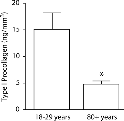
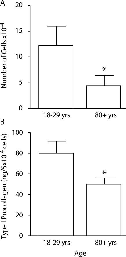
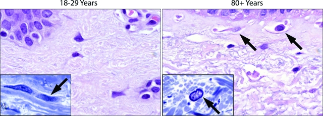
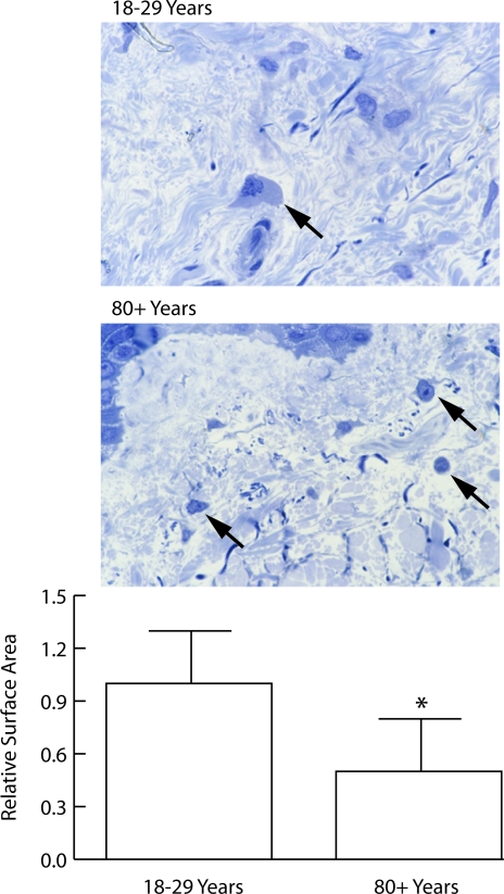
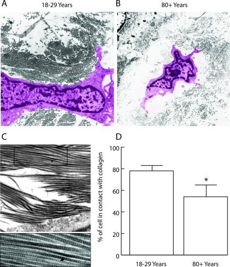
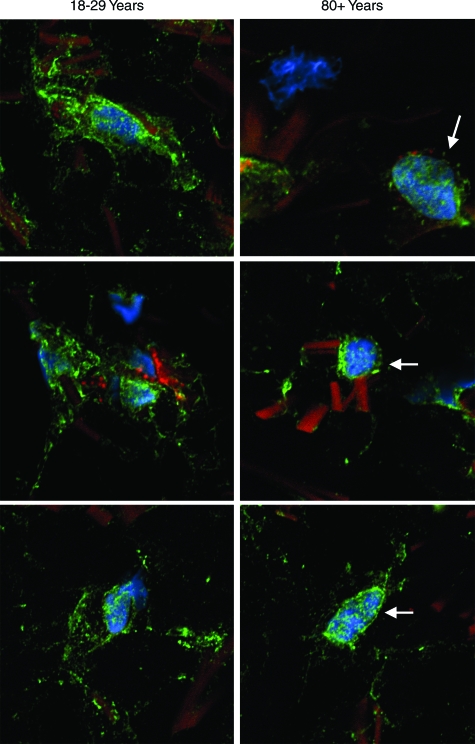
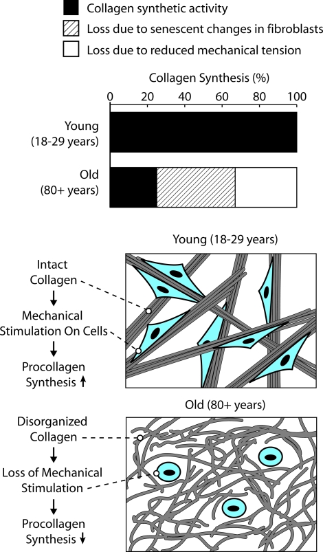
Reduced synthesis of collagen types I and III is characteristic of chronologically aged skin. The present report provides evidence that both cellular fibroblast aging and defective mechanical stimulation in the aged tissue contribute to reduced collagen synthesis. The reduction in collagen synthesis due to fibroblast aging was demonstrated by a lower in vitro production of type I procollagen by dermal fibroblasts isolated from skin of young (18 to 29 years) versus old (80+ years) individuals (82 +/- 16 versus 56 +/- 8 ng/ml; P < 0.05). A reduction in mechanical stimulation in chronologically aged skin was inferred from morphological, ultrastructural, and fluorescence microscopic studies. These studies, comparing dermal sections from young and old individuals, demonstrated a greater percentage of the cell surface attached to collagen fibers (78 +/- 6 versus 58 +/- 8%; P < 0.01) and more extensive cell spreading (1.0 +/- 0.3 vs. 0.5 +/- 0.3; P < 0.05) in young skin compared with old skin. These features are consistent with a lower level of mechanical stimulation on the cells in old versus young skin. Based on the findings presented here, we conclude that reduced collagen synthesis in chronologically aged skin reflects at least two different underlying mechanisms: cellular fibroblast aging and a lower level of mechanical stimulation.


Figures

Figure 1
Figure 1
Type I procollagen production in young and old skin. Values shown are averages ± SEM, based on six young and six old individuals. Statistical significance of the differences between young and old skin was determined using Student’s t-test. *Significance at the P < 0.05 level.
Figure 2
Figure 2
Cellular proliferation and type I procollagen production in monolayer culture. Proliferation (A) and type I procollagen production (B) by fibroblasts isolated from young and old skin. Values shown are averages ± SEM, based on 26 fibroblast isolates from eight young individuals and 37 isolates from eight old individuals. Statistical significance of the differences between isolates from young and old skin was determined using Student’s t-test. *Significance at the P < 0.05 level.
Figure 3
Figure 3
Histological features of sun-protected skin from young and old individuals as observed in 5-μm hematoxylin and eosin-stained sections from formalin-fixed tissue (main frames) and in 1-μm toluidine blue-stained sections from glutaraldehyde-fixed, plastic-embedded tissue (insets). Thick fiber bundles are present throughout the upper dermis of sun-protected young skin. Inset: Some fibroblasts can be seen oriented in the plane of the fiber bundles. In the old skin sample, the bundles have been replaced with thin, disorganized fibers. There is more open space in the dermis. Interstitial cells are round or oblong, and some are surrounded by open space (arrows). Inset: Fibroblast orientation (arrow) is not evident. Hematoxylin and eosin-stained sections, ×490; toluidine blue-stained sections (insets) ×980.
Figure 4
Figure 4
Shape of fibroblasts in the papillary dermis of sun-protected hip skin from young and old individuals (1-μm toluidine blue-stained sections from glutaraldehyde-fixed, plastic-embedded tissue). Top panel: Cells in young skin are flattened, and cytoplasm and nucleus are visible (arrow). Cells are embedded in matrix. Cells in old skin appear round, and only the nucleus and a small amount of cytoplasm are visible (arrows). Bottom panel: Surface area measurements were made quantitatively as described in Materials and Methods. Values represent mean cross-sectional surface area ± SEM, based on 160 cells in sun-protected skin from six young individuals and 57 cells in sun-protected skin from six old individuals. Statistical significance was determined using Student’s t-test (two-tailed). *P < 0.01 (magnification, ×240).
Figure 5
Figure 5
Ultrastructural appearance of dermal fibroblasts in healthy sun-protected hip skin from young and old individuals. A and B: The cell from the section of young skin (A) is flattened and well spread. The cell is in contact with collagen fibers over a high percentage of its surface. The cell in the old skin sample (B) is round and is in contact with collagen polymer over a smaller portion of its surface. There is more open space surrounding the cell. (The computer-generated coloring of the cells was done to aid in the demarcation of cells from extracellular material [magnification ×2050]). C: A high magnification (×3500) of old skin showing the striations in the fibers (typical of collagen). D: Quantification of contact between cells and collagen fibers. Values represent the percentage of the cell boundary in contact with collagen fibers ± SEM (P = 0.01; two-tailed Student’s t-test). Measurements are based on 33 cells in sections of healthy skin from six young individuals and 38 cells in sections from six old individuals.
Figure 6
Figure 6
Adhesion-site protein expression in sections of healthy sun-protected hip skin from young and old individuals. Tissue sections (OCT-embedded frozen) were stained using antibody to vinculin and concomitantly with phalloidin (actin stain) and DAPI (nuclear stain) as described in Materials and Methods. After staining, cells were examined by confocal fluorescence microscopy. Cells are identified by their blue (DAPI)-stained nuclei. Bright green punctate fluorescence identifies vinculin. In the 18- to 29-year-old skin samples, vinculin can be seen at a distance from the nucleus, and in many areas, the vinculin appears to be in close apposition to collagen fibers. In the 80+-year-old skin, blue-stained nuclei are apparent, but there is less vinculin than in the young skin samples. Where intense focal staining is evident, it is surrounding the nucleus (arrows). Away from the nucleus, staining is more diffuse than seen in cells from young skin. The sections presented are representative of young and old sun-protected skin from six individuals, respectively. In both young and old skin, collagen fibers are apparent by their dull orange fluorescence (magnification, ×1200).
Figure 7
Figure 7
Schematic representation of mechanisms underlying reduced collagen synthesis in aged skin.

References

    1. Smith JG, Davidson EA, Clark WM. Alterations in human dermal connective tissue with age and chronic sun damage. J Invest Dermatol. 1962;39:347–356. - PubMed
    1. Lavker RM. Structural alterations in exposed and unexposed aged skin. J Invest Dermatol. 1979;73:559–566. - PubMed
    1. Pieraggi MT, Julian M, Bouissou H. Fibroblast changes in cutaneous aging. Virchows Arch A Pathol Anat Histopathol. 1984;402:275–287. - PubMed
    1. Marks R. London: Martin Dunitz,; Sun-Damaged Skin. 1992
    1. Lavker RM. Gilchrest BA, editor. Cambridge, MA: Blackwell Science,; Cutaneous agingchronologic versus photoaging. Photoaging. 1995:pp 123–135.

Publication types